Which side of the pancreas: functions, structure
Each organ is an integral instrument of a complex mechanism. It has a unique location and role. To successfully interact with your body, it is important to know the location of the internal "helpers". This will help to take the necessary measures in a timely manner in the event of any pathology. To understand from which side the pancreas is, one should study the structure of the gastrointestinal tract. For a quick determination, draw an imaginary line 10 cm above the navel, closer to the left side.

Which side is the pancreas
Content
- 1 Pancreatic functions
- 2 Pancreas: curious facts
-
3 The structure of the pancreas
- 3.1 Head
- 3.2 Body
- 3.3 Tail
- 3.4 Duct
- 3.5 Microscopic structure
-
4 Location of the pancreas
- 4.1 Video - The pancreas: where is it, how it hurts, symptoms
Pancreatic functions
The second after the liver in size, but not in importance, the gland is considered an active participant in the digestion process. It is endowed with several essential functions:
- exocrine;
- endocrine.

The pancreas is located in the abdominal area next to the stomach
The exocrine "mission" is to produce juice containing enzymes for processing food. Endocrine - in the production of hormones and metabolic control.
Table 1. Pancreatic functions
| Function | Description |
|---|---|
| Exocrine | Every day, the gland produces from 500 to 1000 ml of liquid, the composition of which is rich in water, pro-enzymes and salts. When the food lump enters the duodenum, the process of producing hormones that affect the digestive protein organic matter is triggered. Hydrochloric acid found in gastric juice is the most powerful activator of pancreatic secretion. |
| Endocrine | Insulin and glucagon production. These hormones appear due to the work of cells accumulated in the tail of the gland. They are localized between the lobules of the organ, which do not contain the excretory ducts - the islets of Langerance, in scientific terms. |
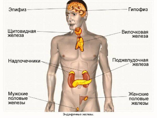
Endocrine glands
The food in the body provokes the production of digestive secretions by the gland. Since the enzymes contained in the secretion are able to process their own tissues, they are produced in an inactive state. Only when they enter the intestines, the process of their activation is started, leading to the digestion of the food lump. At the end of the digestion of food, the elements formed during the decay process are absorbed by the circulatory system.
The hormones produced by the gland are heard by every person who does not know medical terminology:
-
Insulin. A unique hormone of its kind that can lower blood sugar levels. In a healthy body, an increase in glucose levels triggers an immediate release of insulin. It is initially produced in an inactive form. As it enters the bloodstream, a chain of biochemical reactions acts as a trigger. The activation of the hormone leads to the utilization of glucose and other sugar compounds. In the presence of appropriate pathologies, the production of this important element decreases, which is fraught with hyperglycemia and the occurrence of diabetes mellitus.

Insulin functions
- Glucagon. Unlike "spontaneous" insulin, the rhythm of the production of this hormone is rather monotonous. He is constantly busy releasing glucose from complex compounds, which contributes to the rise in blood sugar. For optimal functioning of the central nervous system and other systems, it is necessary to maintain the proper level of this element.

Insulin and glucagon
Sometimes C-peptide is classified as a hormone produced by the pancreas. However, it is more correct to classify it as a protein fragment, since it does not have biological activity.
Pancreas: curious facts
Historically, this multifunctional organ has received less attention than, for example, the heart, lungs or intestines. A few centuries ago, the importance of the gland for the body was underestimated or completely leveled out.

Pancreas location
Interesting information:
- "Abdominal salivary gland". This is the definition that the greatest minds of medicine called this organ several centuries ago. If you dig deep into the centuries, you can find out that the pancreas was considered a pillow for the stomach or "liver finger". And at the dawn of the last millennium, doctors saw in it only a muscle or a lymph node. Confusion over differentiation has led to the gland officially becoming an "unnamed organ." XVII century. made the necessary adjustments: the French physiologist C. Bernard dotted the "i" and figured out its purpose. Information about the role of the gland in the digestive process gradually accumulated and in the last century, scientists finally guessed that the pancreas produces not saliva, but juice.
- Pancreatic juice - a colorless liquid, the pH of which is from 7.5 to 8.8. Under its influence, the food lump in the intestine breaks down into products that are absorbed by the body.
- Characteristics of the formed organ: length - from 14 to 18 cm, width - from 3 to 9 cm, thickness - from 2 to 3 cm, weight - from 70 to 80 g.
- The enzymatic composition of the secreted fluid is determined by the type of food consumed. Carbohydrates, for example, interfere with the secretion of amylase, while fatty foods promote lipase production.
-
The organ begins to form at the 3rd week of fetal development., and at 3 months the process of creating the ducts starts. In an adult, the pancreas is fixed in the peritoneum, while in infants it is quite mobile. The final "maturation" ends upon reaching 6 years of age.

Pancreatic juice composition
The most important role of the organ is to transform food into an energy resource necessary for the body. Minor malfunctions in the functioning of the pancreas lead to a disruption in the production of hormones and enzymes, which negatively affects metabolic and digestive processes and the functioning of the thyroid gland.
The structure of the pancreas
The organ belongs to the category of tubular-alveolar glands. The main anatomical elements are glandular and connective tissues. The shape of the gland is oblong, the color is gray-pink. From the outside, it is wrapped in a connective tissue capsule of fine texture.
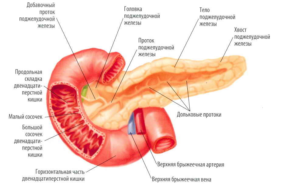
Pancreas
The pancreas consists of several fragments:
- head;
- body;
- tail.
In the area between the head and the body, there is a narrow isthmus called the neck.

Pancreatic ducts
There are no delineated boundaries between departments. The division principle is based on the location of adjacent organs or formations relative to the gland. Any fragment includes several parts, subdivided into smaller ones. They are equipped with excretory ducts flowing into interlobular.
Head
The first section of the pancreas is the widest in diameter. It is located on the right side of the lumbar vertebrae. In its lower part, the head bends, resembling a hook in shape.

The shape of the body of the pancreas resembles a prism.
Peculiarities:
- Almost closely adjacent to the duodenum, forming a small loop.
- Between the head and the lower part of the duodenum there is a small gap in which the bile duct is located.
- The renal artery and the vena cava are connected to the back of this section.
- The abdominal aorta is adjacent to the neck of the gland.
- The front part is "covered" by the abdominal wall, and in the central part it intersects with the colon.
- The location of the head coincides with the area of the small intestine.
The posterior fragment of the neck is provided with a notch in which the portal vein is located.
Body
Localized in the area of the first vertebral vertebra. The right side of the case is raised upward in a slight bend, while the left one looks down. On the left, a kidney adjoins it. The anterior part is equipped with an omental tubercle located in the region of the lesser gastric curvature. Behind is the aorta and renal vein. The lower section is in contact with the small intestine.

Small intestine
Tail
Located on the left side. The greatest functional load falls on this part of the gland. It begins in the region of the posterior surface of the peritoneal wall and "moves" upward with a slight slope to the left. The sizes of the appendix are from 16 to 22 mm. It resembles a hammer in shape. The glandular tissue and islets of Langerhans are the main constituents of this site. At the bottom of the tail is the bend of the colon, and at the back is the left kidney.

Kidney structure
Duct
A duct is located along the entire perimeter of the gland, localized in its front part. It flows smoothly into smaller ducts. Through these channels, pancreatic juice is sent to the duodenum. In the right part of the head, it is combined with the bile duct.

Location of the duodenum
During a person's life, the pancreas undergoes transformations. Both the volume of the organ and its location change, as well as the functional range. It is noticed that upon reaching 50 years of age, the activity of the islets, which take an active part in the production of hormones, decreases.
Microscopic structure
The structure of the tissues of the gland is equally important. Several types of cells are adjacent to the connective tissue - the stroma. It contains the excretory ducts and blood vessels. It is in this layer that the withdrawal of the pancreatic secretion occurs.
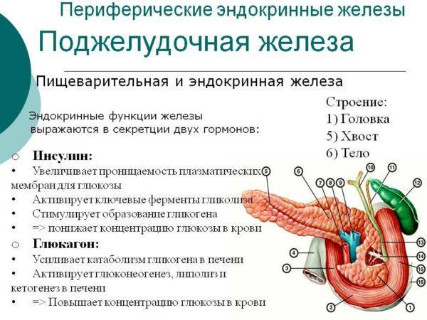
Endocrine functions of the pancreas
Pancreatic cells are divided into 2 types:
- Endocrine. They produce hormones and transport them to the vessels. They are combined into groups - islets of Langegrands, in which alpha cells produce glucagon, beta - insulin,
- Exocrine. Produces enzymes that aid the digestion process. Each cell contains a granule filled with active elements. They are united by a common duct ending in a duodenal papilla.

The structure of the exocrine pancreas
The pancreas includes many small glands, made up of a single layer of cells. They surround the central lumen, into which pancreatic juice is directed.
Location of the pancreas
Aristotle was the first to shed light on the existence of this organ. More extensive data on its anatomical structure were later given by R. de Graaf is a Dutch physiologist. However, only in the last century, thanks to the capabilities of abdominal ultrasound, it became possible to examine the pancreas and determine its functionality.

Depending on the position of the human body, the location of the pancreas changes.
The peculiarities of the location of the gland are contained in the name of the organ. If a person is in a standing position, the pancreas is located behind the stomach walls. Directly under the stomach, it is only when taking a lying position. The peritoneum reliably protects the pancreas by wrapping its lower and anterior parts. The back part is not in contact with the lining of the abdominal cavity. The spine and lumbar muscles protect the gland from external damage.
It is localized in the region of the first and second lumbar vertebrae. The axis of the organ is at right angles with a slight shift to the left in relation to the axis of the spinal column. In front, the pancreas is placed a few fingers above the navel.
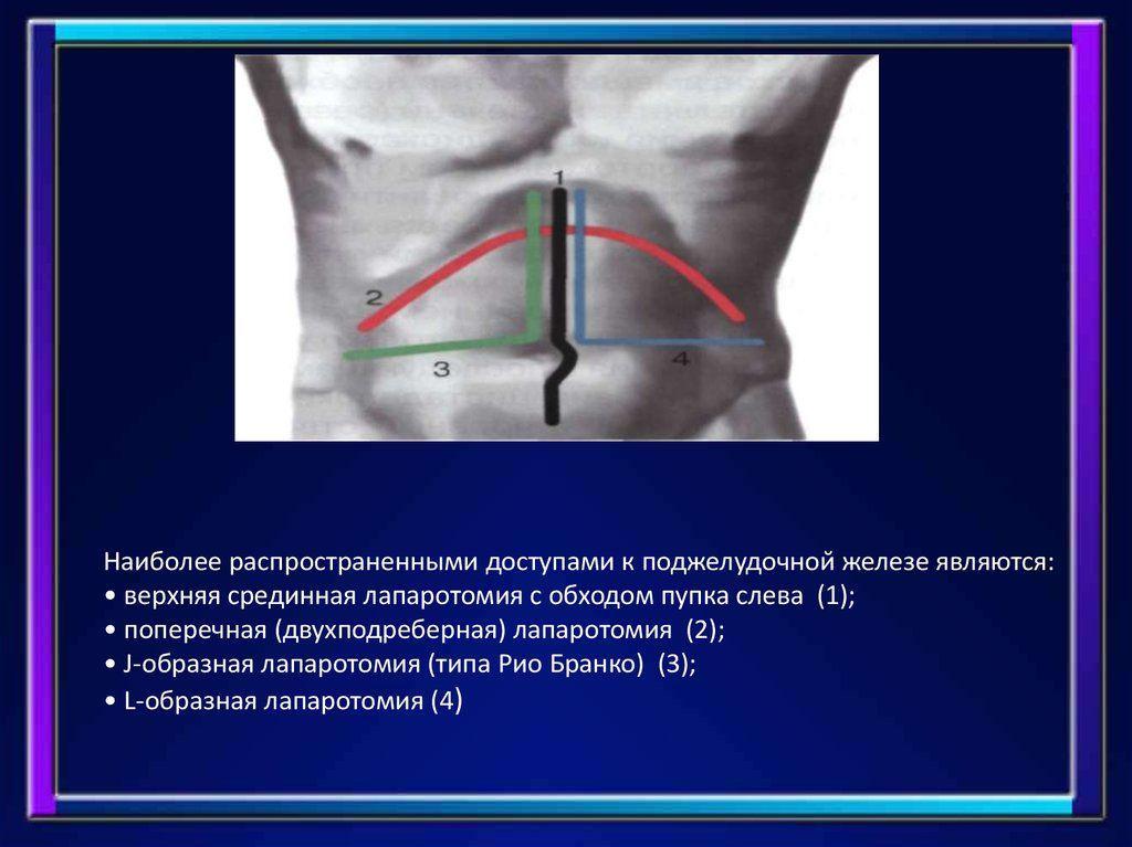
The most common access to the pancreas
In relation to the anterior part of the abdominal zone, the gland is located as follows:
- head - left hypochondrium;
- tail - right hypochondrium;
- body - epigastrium.
To visually determine the location of the organ, you can measure the distance between the end of the chest and the navel and calculate the middle. It is in this area that most of the pancreas is concentrated. The lower part of the organ is 5 cm above the navel, and the upper point is 10 cm.
Information about the projection zones will enable the patient to draw a preliminary conclusion about the source of the discomfort in the event of any pathologies. If the organ is inflamed, the pain is concentrated mainly in the epigastric region, sometimes radiating to the hypochondrium. In acute complications, painful manifestations affect the entire anterior part of the abdominal cavity. Only a specialist can determine the exact source of pain.

Pancreas ultrasound
The deep location of the organ in the peritoneum significantly complicates the diagnosis of pancreatic diseases. An accurate picture of the condition of the gland can be obtained during an ultrasound examination. It allows you to set its size, study the shape and condition of the shell. These data will help to make the correct diagnosis.