Budd-Chiari Syndrome: what is it, causes, symptoms, treatment, prognosis
Content
- general information
- Pathogenesis
- Causes
- Signs and symptoms
- Complications
- Diagnostics
- Treatment
- Surgical therapy
- Forecast
general information
Budd-Chiari Syndrome or veno-occlusive liver disease characterized by obstruction of the hepatic veins and retention of blood in the liver.
After blood enters the liver, it returns to the systemic circulation through the hepatic veins and then into the inferior vena cava, a large vessel that returns blood back to the heart. In veno-occlusive diseases, this blood circulation is partially blocked. The immediate result is liver congestion - retention of blood flowing to the liver. The liver becomes noticeably painful, increases in volume and causes discomfort.
This condition causes the accumulation of fluid in the abdominal cavity -ascites, one of the most common symptoms of Budd-Chiari syndrome.
If the obstruction is severe and affects the vessels that carry blood from the rest of the lower body to the heart - the inferior vena cava, peripheral edema.
Another less noticeable, but no less serious sign is
portal hypertension. It is characterized by increased pressure in the portal veins due to a blockage of blood that must flow from the liver. Blocked veins cause other branch veins to expand - phlebeurysm. The retained blood can return to the heart through the gastroesophageal veins. They are fragile and cannot cope with high blood pressure and burst, causing bleeding in the gastrointestinal cycle from varicose veins.The condition occurs mainly in people with thrombotic diathesis, including myeloproliferative disorders:
- primary polycythemia (polycythemia vera);
- paroxysmal nocturnal hemoglobinuria (PNH);
- tumors;
- chronic inflammatory diseases;
- coagulation disorders;
- infections.
In the short term, for symptomatic therapy may be prescribed drug treatment. If medication alone is used, it will lead to death in 83% of cases. Therapy includes the administration of drugs to control ascites, anticoagulant, antithrombotic and angioplastic therapy.
Surgical therapy in combination with drug therapy, increases patient survival. It consists of surgical decompression or transjugular intrahepatic portosystemic shunting (TIPS). If decompensated cirrhosis occurs, liver transplantation is possible.
Forecast negative for untreated patients. As a result, progressive liver failure within 3 months to 3 years from the date of diagnosis, a person dies. The 5-year survival rate after portosystemic shunting is 38-87%. After liver transplantation, the 5-year survival rate is 70%.
Pathogenesis
Intrahepatic vein obstruction in Budd-Chiari syndrome leads to congestive hepatopathy. It occurs as a result of blockage of veins of small or large caliber, with secondary congestion of blood in the liver, as blood enters the liver but cannot exit. As a result of microvascular ischemia due to accumulation of blood, hepatocellular damage occurs.
Blood blocked in the liver returns to the heart through smaller venous bypass networks that cannot handle the amount and pressure of hepatic blood. These include the esophageal and gastric venous networks, which cause dilation of blood vessels and the formation of varicose veins. Varicose veins will eventually rupture the vein and cause severe bleeding in the digestive tract.
Portal hypertension and impaired liver protein synthesis will provoke the accumulation of ascites fluid in the abdominal cavity. The kidney also contributes to this pathological condition by retaining water and salt, activating the renin-angiotensin-aldosterone system.
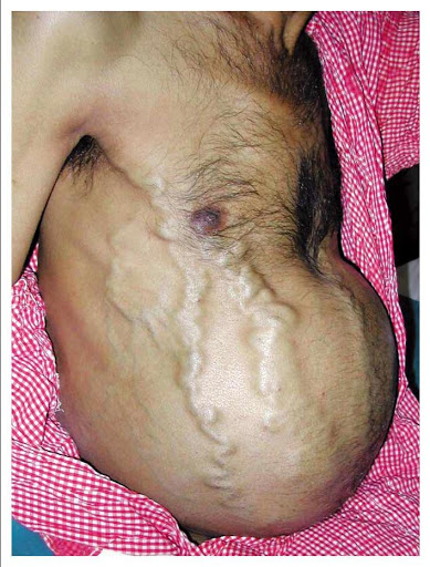
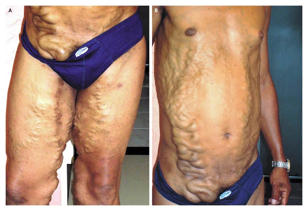
Obstruction can also spread to the inferior vena cava, causing noticeable dilated veins in the abdomen.
Liver stasis and blood congestion lead to hypoxia and the release of free oxygen radicals, which will destroy the hepatocytes. These mechanisms will result in liver necrosis in the central lobular areas with progressive central lobular fibrosis, regenerative nodular hyperplasia and cirrhosis of the liver.
Causes
Most patients with the syndrome have thrombotic disorders. The causes of Budd-Chiari syndrome are as follows:
-
Hematological disorders:
- polycythemia vera, paroxysmal nocturnal hemoglobinuria;
- pathological myeloproliferative conditions, antiphospholipid syndrome;
- essential thrombocytosis.
-
Hereditary thrombotic diathesis:
- deficiency of C and S protein;
- deficiency of antithrombin III;
- deficiency of factor V Leiden.
-
Chronic infections:
- hydatid disease (cystic echinococcosis);
- aspergillosis;
- amebic abscess;
- syphilis;
- tuberculosis.
-
Chronic inflammatory diseases:
- Behcet's disease;
- inflammatory bowel disease;
- sarcoidosis;
- systemic lupus erythematosus;
- Sjogren's syndrome;
- mixed connective tissue disease.
-
Tumors:
- hepatocellular carcinoma (liver cancer);
- renal cell carcinoma;
- leiomyosarcoma
- kidney cancer;
- Wilms tumor (nephroblastoma);
- myxoma of the right atrium.
Have also been associated with Budd-Chiari syndrome oral contraceptive use, pregnancy and postpartum conditions.
Signs and symptoms
The clinical picture of Budd-Chiari syndrome depends on how quickly the occlusion of the hepatic veins expands and whether a collateral venous network has been developed to decompress the liver sinusoids.
The syndrome can be classified as fulminant, acute, subacute, or chronic.
In patients with fulminant form of the syndrome, hepatic encephalopathy eight weeks after development jaundice. Acute patients have short-term symptoms, intractable ascites and liver necrosis without the formation of venous collaterals (bypass paths of blood flow).
The subacute form is the most common and has a more insidious course. Ascites and hepatic necrosis may be minimal because the hepatic sinusoids are decompressed by portal venous circulation. When Budd-Chiari syndrome is acute, thrombosis of all major hepatic veins is common compared to the subacute form, in which it is present in only one third of patients.
The chronic form is a complication of liver cirrhosis.
Stomach ache, hepatomegaly (enlarged liver) and ascites present in almost all patients with Budd-Chiari syndrome. However, asymptomatic patients have also been described. Nausea, vomiting, and mild jaundice most often occur in a fulminant and acute form, while splenomegaly (enlarged spleen) andesogastric varicose veins can be found in chronic forms. When the inferior vena cava is blocked, dilated venous collaterals are present on the flanks next toswelling of the legs.
With the disease, fluid may build up in the abdomen, causing ascites or peripheral leg edema. In patients with hematosis, varicose veins may rupture and bleed. If cirrhosis of the liver develops, it can lead to liver failure with impaired brain function - liver encephalopathy, that leads to confusion and coma.
Sometimes symptoms appear suddenly, as with occlusion of the hepatic veins during during pregnancy. Patients feel fatigue, and the liver increases in volume. Appear pain in the right hypochondrium. Additional symptoms include vomiting, jaundice.
Tricuspid regurgitation, constrictive pericarditis and myxoma of the right atrium may have different manifestations than with Budd-Chiari syndrome. Absence hepato-jugular reflux when abdominal pressure is applied, it excludes the cardiac cause of ascites.
Complications
List of complications arising from Budd-Chiari syndrome:
- hepatic encephalopathy;
- bleeding from varicose veins;
- hepatorenal syndrome;
- portal hypertension;
- complications of a secondary hypercoagulant state.
Diagnostics
Signs that lead a doctor to suspect a patient with Budd-Chiari syndrome include hepatomegaly, ascites, and high levels of liver enzymes found on blood tests. However, in order to resolve all doubts and find out the reasons for the suspicious signs and symptoms, it is necessary to conduct the following studies:
- Doppler ultrasound examination of the liver with a sensitivity and specificity of more than 85% is the preferred liver test for suspected Budd-Chiari syndrome. There are necrotic areas of the liver, blood clots.
- Magnetic resonance imaging (MRI) helps to highlight the venous circulation of the liver. It has a sensitivity and specificity of 90%. It is useful for the study of the inferior vena cava and allows you to differentiate between acute, subacute and chronic forms of the syndrome.
- CT scan (CT) may be useful for determining the anatomy of the venous circulation and for transjugular intrahepatic portosystemic shunting.
- At liver venography blood clots in the hepatic veins can be observed.
- Echocardiography may be required for some patients to investigate tricuspid regurgitation, constrictive pericarditis, or right atrial myxoma.
- Liver biopsy may be required to confirm the diagnosis and detect the development of liver cirrhosis.
Differential diagnosis:
- constrictive pericarditis;
- right-sided heart failure;
- metastatic liver disease;
- alcoholic liver disease;
- granulomatous liver disease.
Treatment
Treatment for Budd-Chiari Syndrome depends on how quickly it develops and how severe the illness is. When symptoms begin suddenly and a blood clot is the cause, fibrinolytic drugs are helpful. Long-term anticoagulant therapy prevents clot enlargement and recurrence.
Anticoagulant agents used include:
- Warfarin - inhibits the synthesis of vitamin K-dependent coagulation factors in the liver.
Fibrinolytic agents:
- Streptokinase - works together with plasminogen, converting it into plasmin. Plasmin breaks down fibrin clots, fibrinogen, and other plasma proteins;
- Urokinase is a direct plasminogen activator that acts on the endogenous fibrinolytic system and converts fibrinogen into plasmin;
- Alteplase is a tissue plasminogen activator used in the treatment of acute myocardial infarction, acute ischemic heart disease and pulmonary embolism; it is safe and effective when given with heparin or aspirin within the first 24 hours after symptom onset.
Ascites to be treated diuretics (diuretics) and limiting water and salt. The most commonly used antidiurics are spironolactone and furosemide. When ascites is energized or does not respond to diuretic therapy, paracentesis and intravenous albumin are needed.
Surgical therapy
If a vein is blocked or ruptured, angioplasty may be done. Transluminal or transhepatic percutaneous angioplasty for localized segments of the occlusive vein or inferior vena cava improves symptoms in more than 70% of patients. The risk of restenosis is high.
If thrombolytic therapy is ineffective, transjugular intrahepatic portosystemic shunting is performed (TVPSh). The placement of this artificial vessel between the hepatic and portal veins is useful in patients with a blocked inferior vena cava, those whose pressure between the portal vein and intrahepatic veins is below 10 mm Hg, and in patients with severe hepatic failure. The procedure is also recommended for patients with acute syndrome who have not been helped by thrombolytic therapy.
In other cases, a blocked vein can be cleared of a blood clot, and then the vein can be a thin rod (stent) is inserted to maintain blood flow.
In severe cases of Budd-Chiari Syndrome, you may need liver transplant. With liver transplant, the survival rate is 95%. The indications are fulminant liver failure, cirrhosis.
Long-term anticoagulant therapy may be required after transplant, although many myeloproliferative disorders have been controlled with aspirin and hydroxyurea.
Forecast
Factors associated with a favorable prognosis are young age at the time of diagnosis, the absence of a large amount of ascites, and a low incidence of serum creatinine. Identification of the cause of Budd-Chiari syndrome using radio images (ultrasound, CT, MRI, etc.) has the greatest impact on the prognosis.
Medication and surgery can extend the life of patients to a rough 8 years, after which liver transplantation is recommended.