Corneal dystrophy: types, symptoms, causes, treatment
Content
- What is corneal dystrophy?
- Signs and symptoms
- Causes
- Affected populations
- Related disorders
- Diagnostics
- Standard treatments
What is corneal dystrophy?
Corneal dystrophy are a group of genetic, often progressive, visual impairments in which foreign materials accumulate in the transparent outer layer of the eye (cornea). In some people, corneal degeneration may not cause symptoms; in others, it can cause significant visual impairment.
Age of onset and specific symptoms vary among different forms of dystrophy. The various forms of the disorder have some of the same symptoms and signs - most forms of corneal dystrophy affect both eyes (bilateral), progressing slowly, not affecting other areas of the body, and tending to appear in families. Most forms are inherited as autosomal dominant traits; some are inherited as autosomal recessive traits.
An international classification of disorders was developed, which takes into account the chromosomal loci of various corneal dystrophies, as well as the responsible genes and their mutations. Traditionally, these disorders have been classified based on their clinical features and the specific layer of the cornea affected. Advances in molecular genetics (such as the identification of specific disease genes) have made it possible to better understand these disorders.
Signs and symptoms
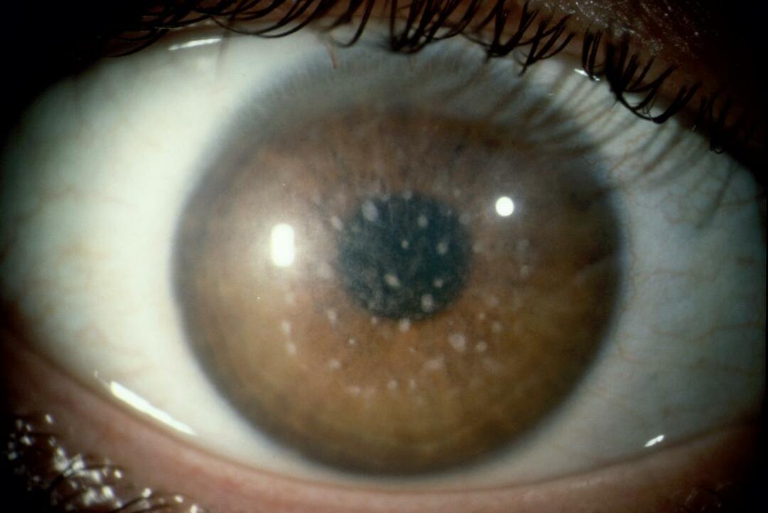
Symptoms of corneal dystrophy result from the accumulation of abnormal material within the cornea, the transparent outer layer of the eye. The cornea (cornea) has two functions - it protects the rest of the eye from dust, germs and other harmful or irritating substances and acts as the outer lens of the eye, bending the incoming light onto the inner lens, where the light is then directed to the retina (the membrane layer of light-sensitive cells at the back eyes). The retina converts light into images, which are then transmitted to the brain. The cornea must remain transparent so that incoming light can be focused.
The cornea is made up of five distinct layers:
- epithelium, the outermost protective layer of the cornea;
- Bowman's membrane (Bowman's membrane), this is the second extremely durable layer that protects the eyes;
- the stroma, the thickest layer of the cornea, made up of water, collagen fibers and other connective tissue components that give the cornea strength, elasticity and transparency;
- Descemet's sheath, a thin, strong inner layer that also acts as a protective layer;
- endothelium, the innermost layer of specialized cells that drain excess water from the cornea.
Corneal dystrophies are characterized by the accumulation of foreign material in one or more of the five layers of the cornea. Such material can cause loss of transparency in the cornea, which can result in loss or impairment of vision.
A symptom common to many forms of corneal dystrophy is recurrent corneal erosion, in which the outer layer (epithelium) does not adhere properly to the eye. Periodic corneal erosion can cause discomfort or severe pain, abnormal sensitivity to light (photophobia), sensation of a foreign body in the eye (such as dirt, dust, or eyelash) and blurred vision.
- Anterior or superficial corneal dystrophy.
These dystrophies affect the outer layers of the cornea, including the epithelium, epithelial basement membrane (a thin membrane that separates epithelial cells from the underlying tissue) and Bowman's membrane.
- Dystrophy of the basement membrane of the epithelium.
This form of the disorder is characterized by the development of very tiny dots (microcysts), gray areas that aggregates resemble the outlines of countries on a map, or thin lines that resemble fingerprints on the epithelial layer of the cornea. For most people, this form of the disease is asymptomatic. In some cases, symptoms may include recurrent erosion and blurred vision, which seems to affect 10% of people. Abnormal sensitivity to light (photophobia) and a sensation of foreign material in the eye may also occur. Dystrophy of epithelial basement membranes is a common form of corneal dystrophy and is also known as Kogan microcystic epithelial dystrophy.
- Youthful epithelial dystrophy of Mesmann.
This extremely rare form of corneal dystrophy affects the epithelial layer of the cornea. It is characterized by the development of clusters of many small, clear cysts. The cysts are about the same size. Affected persons may experience mild irritation and a slight decrease in clarity (acuity) of vision. Also, with this form of dystrophy, sensitivity to light (photophobia) and excessive formation of tears (lacrimation) can occur. Clouding (opacity) of the cornea is rare but can develop in some older people.
- Epithelial dystrophy of the Lish cornea.
This rare form of the disease is characterized by clusters of multiple, tiny cysts or lesions that may appear as stripes that are curved or spiral (curled) in appearance. In some cases, sick people do not have any symptoms. Some people may have blurred vision, blurred vision, and double visionaffecting only one eye (monocular diplopia).
- Reis - Bucklers corneal dystrophy.
This form affects Bowman's membrane and is characterized by cloudiness (opacity) and progressive scarring of the membrane. During the first decade of life, affected individuals may initially develop recurrent erosions causing significant pain. Periodic erosion may eventually stabilize as the affected people grow older. Additional symptoms may appear, including abnormal sensitivity to light (photophobia), foreign body sensation in the eye, and a marked decrease in visual clarity, often by age 20. Reis-Bucklers corneal dystrophy is also known as type I Bowman's membrane dystrophy.
- Thiel-Behnke corneal dystrophy.
This form of the disease also affects Bowman's membrane and can be extremely difficult to distinguish from Reis-Bucklers dystrophy. Disorders affecting the cornea may resemble a honeycomb. Recurrent corneal erosions begin in childhood, but visual acuity is not affected until later in life. Pain and photophobia may also occur. Thiel-Behnke corneal dystrophy is also known as Bowman's membrane dystrophy type II.
- Stromal corneal dystrophy.
These corneal dystrophies affect the stromal or central layer of the cornea. Some of these disorders can progress to affect other layers of the cornea.
- Type I granular corneal degeneration.
This form of the disease is characterized by the development of small particles (granules) that collectively resemble breadcrumbs, usually during the second or third decade of life. These lesions grow slowly, eventually coalescing (coalescing) into larger lesions. People can develop periodic erosion. Although vision loss is usually invisible in the early stages of the disease, visual acuity may decrease by the fourth or fifth decade. Some people may have photophobia. As a result of periodic corneal erosion, eye pain can occur.
- Type II granular corneal degeneration.
In type II granular corneal dystrophy, also known as Avellino corneal dystrophy, lesions develop on the stroma, usually beginning in the first or second decade of life. Opacities in the cornea sometimes resemble a mixture of type 1 granular corneal dystrophy and ethmoid corneal lesions. below). As patients age, lesions can become larger, more visible and cover the entire stromal layer. In some older people, the clarity of vision is reduced due to clouding of the cornea. In some cases, periodic erosion may develop.
- Lattice corneal dystrophy.
Lattice corneal dystrophy is a common form of stromal dystrophy, and two main variants have been identified. They are characterized by the development of lesions that form branching lines resembling cracked glass or crossed, overlapping lattice patterns. Lattice type I dystrophy and its varieties usually appear by the end of the first decade. Periodic erosions (which can be painful) often precede characteristic changes in the stroma. Victims may have decreased visual acuity and photophobia.
Type II lattice dystrophy is classified as corneal dystrophy, but occurs as part of a larger disorder. called familial amyloid polyneuropathy type IV, which is more serious than horny disease shell.
- Gelatinous drop-shaped corneal dystrophy.
Gelatinous teardrop dystrophy, also known as familial subepithelial dystrophy, develops in humans during the first decades of life and is characterized by loss of vision, abnormal sensitivity to light, excessive lacrimation and sensation of foreign bodies in the eye. Gelatinous masses of amyloid, a type of protein, accumulate under the epithelium of the cornea and make the cornea opaque and gradually impair vision.
- Spotted corneal dystrophy.
People with this form of the disease are born with clear corneas, but over time, people develop stromal opacities, usually between the ages of 3 and 9. The progression of the lesions results in decreased visual acuity and irritation early in life. In some cases, significant vision loss may occur by the second decade. Severe vision loss can develop by the third or fourth decade. Sometimes painful recurrent erosions occur, but they are less common than with other dystrophies that affect the stroma. Spotted corneal dystrophy is also known as type II Grenouve corneal dystrophy.
- Crystalline dystrophy of the Schneider cornea.
This form of the disease usually develops during the second decade of life, but can develop as early as the first year of life. Affected people develop clouding of the cornea due to accumulation of fat or cholesterol in the stroma, which ultimately causes blurred vision. Affected people have vision impairment that is exacerbated by bright light.
— Fuchs endothelial dystrophy.
This form of the disease usually develops in middle age, although initially there may be no symptoms. Fuchs' dystrophy is characterized by problems with tiny cells called "pumping" cells (endothelium) in the innermost layer of the cornea. These cells usually pump water out of the eye. With Fuchs's dystrophy, these cells are destroyed ("die off"), and the cornea fills with water and swells. Vision deteriorates, edema appears, which intensifies in the morning, but gradually improves during the day. Tiny blisters form on the cornea, which eventually rupture and cause severe pain. Affected people may also have a gritty sensation inside the eye (foreign body), be highly sensitive to light, and see glare or halos when looking at light. As the disease progresses, vision no longer improves during the day and significant vision loss may occur, possibly requiring a cornea transplant.
- Posterior polymorphic corneal dystrophies.
This unusual form of dystrophy may be present at birth (with corneal opacity) or later in life and is characterized by lesions affecting the endothelium. Most people do not develop symptoms. The effects on the cornea can be slowly progressive. Both eyes are usually affected, but one eye may be more affected than the other (asymmetric). In severe cases, people with posterior polymorphic dystrophy may develop stromal edema, photophobia, decreased vision, and foreign body sensation in the eye. In rare cases, increased pressure in the eye (intraocular pressure) may occur.
- Congenital hereditary corneal dystrophy.
There are two types of congenital hereditary corneal dystrophy: one is inherited as an autosomal dominant trait, and the other is inherited as an autosomal recessive trait. The autosomal dominant form (type I) is characterized by corneal edema, pain. At birth, the cornea is clean, but becomes cloudy in early childhood. The autosomal recessive form (type II) is characterized by corneal edema and corneal opacity at birth. Rapid, shaky eye movements (nystagmus). The recessive form is more common than the dominant one.
Causes
Most cases of corneal dystrophy are inherited as an autosomal dominant trait with variable expressivity. Genetic diseases are defined by a combination of genes for a specific trait, which are found on chromosomes received from the father and mother.
Dominant genetic disorders occur when only one copy of an abnormal gene is needed for a disease to appear. The abnormal gene can be inherited from either parent or be the result of a new mutation (gene change) in the affected person. The risk of passing the abnormal gene from the affected parent to the offspring is 50% with every pregnancy, regardless of the sex of the child.
Variable expressivity means that some people who inherit the same dominant disorder gene may not show the same symptoms.
Dystrophy of the basement membrane of the epithelium, Reis - Bucklers, Thiel - Behnke, Mesmann, Schneider, ethmoid type I and II, granular type I, granular type II (Avellino), congenital hereditary corneal dystrophy of type I and posterior polymorphic forms are autosomal dominant inheritance. Fuchs' dystrophy can have autosomal dominant inheritance in some cases; in others, it may occur spontaneously for no apparent reason (sporadically). Spotted dystrophy and congenital hereditary corneal dystrophy of type II have an autosomal recessive inheritance.
Recessive genetic disorders occur when a person inherits the same abnormal gene for one trait from each parent. If a person receives one normal gene and one gene for the disease, the person will carry the disease but usually show no symptoms. The risk of both carrier parents passing on both defective genes and therefore infecting the baby is 25% with each pregnancy. The risk of giving birth to a carrier child, like the parents, is 50% with each pregnancy. The chance for a child to receive normal genes from both parents and be genetically normal for this particular trait is 25%. The risk is the same for men and women.
Scientists have determined that some corneal dystrophies are due to abnormalities or changes (mutations) of the transforming growth factor beta-1 (TGF-B1) gene located on the long arm (q) of chromosome 5 (5q31). Chromosomes are present in the nucleus of every person's cells and carry genetic information. The cells of the human body usually have 46 chromosomes. Pairs of human chromosomes are numbered 1 through 22, and the sex chromosomes are labeled X and Y. Men have one X chromosome and one Y chromosome, while women have two X chromosomes. Each chromosome has a short arm, labeled "p", and a long arm, labeled "q". Chromosomes are further subdivided into many numbered bands. For example, "chromosome 5q31" refers to lane 31 on the long arm of chromosome 5.
Various epithelial dystrophies, Reis-Bucklers, Thiel-Behnke, granular types I and II, and latticed dystrophy type I have been associated with the transforming growth factor beta (TGFB1) gene. These forms of the disorder develop due to various mutations in this gene, which was previously known as the human Beta IG H3 gene (BIGH3). The TGFB1 gene contains instructions for creating (coding) a protein known as a protein that causes transformation of growth factor beta (keratoepithelin), which helps the stratum corneum to stay sticky (glued) together. The accumulation of this protein due to the mutated gene causes the symptoms of corneal degeneration associated with this gene.
Mesmann's dystrophy is associated with mutations in two separate genes, one (KTR3) on the long arm of chromosome 17 (17q12) and one (KTR12) on the long arm of chromosome 12 (12q13). These genes contain instructions for making (coding) certain proteins called keratins, which are necessary for the cornea to form properly.
Some cases of Thiel-Behnke dystrophy have been associated with mutations in a gene located on the long arm of chromosome 10 (10q23-q24).
Spotted dystrophy is associated with mutations in the carbohydrate sulfotransferase-6 (CHST6) gene on the long arm of chromosome 16 (16q22). This gene encodes keratan sulfate, a complex sulfated carbohydrate essential for the proper development of cartilage and cornea.
Schneider's dystrophy is associated with mutations in the UBIAD1 gene located on the short arm of chromosome 1 (1p34-q36).
Posterior polymorphic dystrophy has been associated with three different chromosomes. One is on the long arm of chromosome 20 (20p11.2), the other is on chromosome 1 (1p34.3-p32.3) with the COL8A2 gene, and the third is due to a mutation in the TCF8 gene on chromosome 10 (10p11-Q11).
An autosomal recessive form of congenital hereditary corneal dystrophy of the endothelium is caused by mutations in the SLC4A11 gene on chromosome 20 (20p13). The gene for autosomal dominant congenital hereditary endothelial dystrophy has not been identified, but it is located on the short arm of chromosome 20 (20p11.2-q11.20).
Lish's epithelial corneal dystrophy was associated with a gene on the short arm of the X chromosome (Xp23). This form of corneal dystrophy is believed to be inherited as an X-linked dominant trait. X-linked dominant disorders are caused by an abnormal gene on the X chromosome. Men with the abnormal gene are more affected than women.
Affected populations
Corneal dystrophies affect women and men in equal numbers, with the exception of Fuchs's dystrophy, which affects women about four times more often than men. Various forms of the disorder can affect people of all ages. The frequency is unknown. Because some people are asymptomatic (asymptomatic), it is difficult to determine the true incidence of disorders in the general population.
Related disorders
Symptoms of the following disorders may be similar to those of corneal dystrophy. Comparisons can be useful for differential diagnosis.
- Keratoconus - non-inflammatory eye disease characterized by progressive changes in the shape of the cornea. The cornea is a thin-walled “domed” transparent layer that forms the anterior portion of the eyeball; it serves as a protective coating and helps focus or bend (refract) light waves onto the retina at the back of the eye. In people with keratoconus, the slow, progressive thinning of the cornea causes it to bulge out or protrude forward as irregular cone-shaped (conical) shape, which leads to blurry vision, increased sensitivity to light and other problems with eyes. Keratoconus often begins during puberty. Although the exact cause of the disease is unknown, scientists point out that genetic factors play a role. In addition, in some cases, keratoconus can occur in connection with a variety of other disorders.
- Bullous keratopathy - an eye condition characterized by edema (inflammation) of the cornea due to abnormal water retention in the cornea. Bullous keratopathy can lead to pain and loss of vision. Small blisters (bullae) can form on the surface of the eye, which can potentially rupture, causing severe pain and infection. Bullous keratopathy can be caused by eye surgery, trauma, and inflammatory diseases of the eye.
Diagnostics
The presence of the disease can be discovered by chance during a routine eye exam. The diagnosis can be confirmed by careful clinical evaluation, a detailed history of the patient, and various investigations such as slit lamp examination, in which a special microscope (slit lamp) allows the doctor to see the eye with a large increase. Some specific corneal dystrophies can be diagnosed with molecular genetic tests before symptoms appear.
Standard treatments
Treatment for corneal dystrophy varies. Individuals who are asymptomatic or have only mild symptoms may not need treatment and may instead be monitored regularly for potential disease progression.
Specific treatments for corneal dystrophy may include eye drops, ointments, lasers, and corneal transplants. Recurrent erosions (common in most dystrophies) can be treated with lubricating eye drops, ointments, antibiotics, or special (soft bandages) contact lenses. If recurrent erosion persists, additional measures such as scraping the cornea or using excimer laser therapy, can eliminate abnormalities from the surface of the cornea (phototherapy keratectomy).
People with significant comorbid symptoms may require a corneal transplant, known as keratoplasty. Corneal transplants have been very successful in treating people with severe dystrophic symptoms. However, there is a risk that lesions will eventually develop in the transplanted (donor) cornea.
Several factors determine which treatments can be used to treat people with the disorder, including the specific type of treatment available. corneal dystrophy, severity of associated symptoms, rate of disease progression, and overall health and quality of life the patient.
Genetic counseling can be beneficial for affected individuals and their families. Other treatments are symptomatic and supportive.