Fanconi anemia: what is it, causes, symptoms, treatment
Content
- What is Fanconi anemia?
- Signs and symptoms
- Causes of Fanconi anemia
- Affected populations
- Diagnostics
- Fanconi anemia treatment
What is Fanconi anemia?
Fanconi anemia (abbr. AF) Is a rare genetic disease in the category of hereditary bone marrow failure syndromes. Half of the patients are diagnosed before the age of 10, and about 10% of the disease is diagnosed already in adulthood.
Affected patients suffer from birth defects such as short stature, abnormal thumbs and / or radial bones, skin pigmentation, small head, small eyes, abnormal structures of the kidneys, and abnormalities of the heart and skeleton.
The disorder is often associated with a progressive deficiency of all bone marrow production of blood cells - erythrocytes, leukocytes, and platelets. Affected people have an increased risk of developing a cancer of the blood-forming cells in the bone marrow, called acute myeloid leukemia (AML), or tumors of the head, neck, skin, gastrointestinal tract, or genital ways.
Fanconi anemia occurs equally in men and women, and across all ethnic groups. It is usually inherited as an autosomal recessive genetic disorder, but X-linked inheritance has also been reported.
There are several subtypes of AF, which are the result of the inheritance of two gene mutations in each of at least 18 different genes. Most of the subtypes have characteristic symptoms and signs. Fanconi anemia is not the same as Fanconi syndrome, a rare kidney disorder.
Signs and symptoms
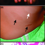

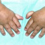
The symptoms of Fanconi anemia vary from person to person. Symptoms identified include a variety of physical abnormalities, bone marrow failure, and an increased risk of developing malignant neoplasms. Physical abnormalities usually manifest in early childhood, but rarely are diagnoses made in adulthood. Problems with blood production often develop between the ages of 6-8.
In most cases, the affected bone marrow occurs in the majority of patients, although the progression and age of onset differ. Patients who live in adulthood can develop head and neck cancers, gynecological and / or gastrointestinal cancers at a much earlier age than the general population, regardless of whether or not they had earlier blood problems.
- Physical abnormalities.
At least 60% of people with AF are born with at least one physical abnormality. The anomaly can include any of the following:
- short stature;
- thumb and hand abnormalities: extra fingers, deformity or absence of thumbs, or an incomplete or missing one of the bones in the forearm;
- anomalies of the skeleton of the hips, spine, or ribs;
- structural kidney problems;
- pigmentation of the skin;
- small head;
- small, crossed, or wide-set eyes;
- low birth weight;
- gastrointestinal problems;
- small reproductive organs in men;
- defects in the tissues separating the chambers of the heart.
People with anemic may experience:
- fatigue;
- increased need for sleep;
- weakness;
- dizziness;
- irritability;
- headaches;
- pale skin color;
- labored breathing;
- cardiac symptoms.
There may be excessive bruising after minimal trauma and spontaneous bleeding from the mucous membranes, especially the gums and nose.
- Insufficiency of the bone marrow.
Bone marrow is a spongy substance found in the center of the long bones of the body. The bone marrow produces specialized cells (hematopoietic stem cells) that grow in eventually develop into erythrocytes (red blood cells), leukocytes (white blood cells), and platelets. Cells enter the bloodstream to travel throughout the body to carry out their specific functions. Red blood cells carry oxygen to the body, white blood cells help fight infections, and platelets allow the body to form clots, stopping bleeding.
Progressive bone marrow failure usually appears by age 10 and is usually accompanied by low platelet counts or low white blood cell counts. By the age of 40-50, the estimated incidence of bone marrow failure as the first serious event is over 50%.
Those affected develop low levels of all cellular elements in the bone marrow - red and white blood cells and platelets, which can lead to the following:
- a low level of circulating red blood cells - anemia;
- low leukocyte count - leukopenia;
- a low level of neutrophils (a type of white blood cell) - neutropenia;
- low platelet count - thrombocytopenia;
- Increased risk of developing malignant neoplasms.
People with AF have a higher risk of developing certain forms of cancer, including acute myeloid leukemia and specific solid tumors, than the general population.
Affected people may have an extremely high risk of developing cancers affecting the head and neck area, gastrointestinal tract, esophagus, or gynecological areas. Most of these are a specific form of cancer known as squamous cell carcinoma. AF patients in whom bone marrow failure is treated with male hormones (called "androgens") are at increased risk of developing liver cancer.
In about 30 percent of cancer-related cases, the development of a malignant tumor precedes the diagnosis of AF.
Causes of Fanconi anemia
Chromosomes in the cells of individuals with Fanconi anemia are unable to repair damage deoxyribonucleic acid (DNA) and thus are easily destroyed and rearranged (chromosome instability). DNA is the carrier of the genetic code, and DNA damage is a common daily occurrence. For most people, DNA damage is repaired. However, in people with AF, fractures and realignments occur more frequently, and their bodies are slow or unable to repair damage.
Mutations in at least 18 genes can cause AF. The proteins encoded by these genes work together in a common pathway called by FAwhich comes into play when DNA damage occurs. The FA pathway directs specific proteins to the site of injury so that DNA can repair itself and continue to be copied (replicated). Eight proteins form a complex known as FA core complex, which activates two genes to form proteins called FANCD2 and FANCI. Activation of these two proteins brings DNA repair proteins to the area of DNA damage.
80-90% of cases of the disease occur due to mutations in one of the three genes, FANCA, FANCC and FANCG. These genes provide instructions for obtaining the components of the FA nucleus complex. Mutations in any of the many genes associated with the major FA complex will render the complex dysfunctional and disrupt the entire FA pathway. Disruption of this pathway leads to an accumulation of DNA damage that can lead to abnormal cell death or abnormal growth. Cell death leads to a decrease in the number of blood cells and physical disorders associated with Fanconi's anemia. Uncontrolled cell growth can lead to the development of acute myeloid leukemia or other cancers.
Most cases of AF are inherited in an autosomal recessive manner. Recessive genetic disorders occur when a person inherits two copies of an abnormal gene for the same trait, one from each parent. If a person inherits one normal gene and one gene for the disease, the person will carry the disease but usually will not show symptoms. The risk for two carrier parents who both pass on the altered gene and infect the baby is 25% with each pregnancy. The risk of conceiving a child who is a carrier as a parent is 50% with each pregnancy. The probability for a child to receive normal genes from both parents is 25%. The risk is the same for men and women.
Parents who are close relatives (brother and sister) are more likely than unrelated parents who have the same abnormal gene, which increases the risk of having children with a recessive genetic disorder.
Mutations in the following genes also cause AF and are inherited in an autosomal recessive manner: BRCA2, BRIP1, FANCB, FANCD2, FANCE, FANCF, FANCI, ERCC4, FANCL, FANCM, PALB2, RAD51C, SLX4 and UBE2T.
Gene FANCB located on the X chromosome and causes less than 1% of all AF cases. This gene is inherited as an X-linked recessive trait.
X-linked genetic disorders are conditions caused by an abnormal gene on the X chromosome and occur primarily in males. Women with an altered gene on one of their X chromosomes are carriers of the disorder. Carrier women usually show no symptoms because women have two X chromosomes and only one carries the altered gene. Men have one X chromosome, which is inherited from their mother, and if a man inherits an X chromosome that contains the altered gene, he will develop the disease. Women with X-related disorder have a 25% chance of having a similar carrier daughter with each pregnancy, 25% chance of having a non-carrier daughter, 25% chance of having a son affected by the disease, and 25% chance of having an unaffected son. If a man with an X-linked disorder is able to reproduce, he will pass on the altered gene to all of his daughters who will be carriers. A man cannot pass on his X-linked gene to his sons, as men always pass on their Y chromosome instead of their X chromosome to male offspring.
Gene mutations RAD51 cause autosomal dominant AF. Dominant genetic disorders occur when only one copy of an abnormal gene is needed to cause a specific disorder. The abnormal gene can be inherited from either parent, or it can be the result of a new mutation (gene change) in the affected person. The risk of passing the abnormal gene from the affected parent to the offspring is 50% for each pregnancy. The risk is the same for men and women. To date, all patients with AF due to gene mutation RAD51 have spontaneous (de novo) a genetic mutation that occurs in an egg or sperm cell. In such situations, the disorder is not inherited from the parents.
Affected populations
The estimated incidence of Fanconi anemia is about 1 in 136,000 births. The disease is more common among Ashkenazi Jews, Roma in Spain and black South Africans.
Diagnostics
The diagnosis of AF is based on careful clinical evaluation, detailed patient history, identification of characteristic features, and various specialized tests.
The definitive test for AF is currently the chromosome rupture test: some of the patient's blood cells are treated in a test tube with a chemical that stitches DNA together. Normal cells are capable of repairing most of the damage and are not severely affected, whereas with disease, cells show marked chromosome destruction. Two chemicals are commonly used for this test: DEB (diepoxybutane) and MMC (mitomycin C). These tests can be performed prenatally on cells from the chorionic villi or from the amniotic fluid.
Blood tests can be performed to determine the levels of red and white blood cells and platelets. X-ray examination can reveal the presence and extent of skeletal malformations and internal structural abnormalities.
Many cases of Fanconi's anemia are not diagnosed at all or are not diagnosed in a timely manner. AF should be suspected and checked for the presence of any child born with the thumb and hand abnormalities described earlier. Anyone who develops aplastic anemia at any age should have an AF test, even if there are no other defects. Any patient who develops at an early age squamous cell carcinoma of the head and neck, gastrointestinal tract or gynecological system with or without the use of tobacco or alcohol, should be examined for AF. Many AF patients show no other abnormalities. An AF test should be performed before considering stem cell transplantation for aplastic anemia or cancer treatment because standard chemotherapy and radiation protocols can be toxic to patients with disease.
Molecular genetic testing is available for all 18 genes associated with AF. Complementation testing is usually done first to determine which gene is mutating. You can then analyze the sequence of the corresponding gene to determine the specific mutation in that gene. If no mutation is identified, gene deletion / duplication analysis is clinically available.
Targeted mutation analysis available for common Ashkenazi Jewish mutation FANCC.
- Clinical research.
To determine the extent of disease in an individual diagnosed with AF, the following tests are recommended, if necessary:
- Ultrasound examination of the kidneys and urinary tract.
- A formal hearing test.
- Development assessment (especially important for toddlers and schoolchildren).
- Contacting an ophthalmologist, otolaryngologist, endocrinologist, hand surgeon, gynecologist (for women, as indicated), gastroenterologist, urologist, dermatologist, ENT surgeon, genetic consultant.
- Evaluation by a hematologist, including complete blood count, fetal hemoglobin and bone marrow aspirate for cell morphology and chromosome studies (cytogenetics), as well as biopsy to determine cellularity.
- HLA typing of individuals, siblings and parents to consider hematopoietic stem cell transplantation.
- Complete blood typing.
- Blood chemistry (assessment of the condition of the liver, kidneys, thyroid gland, lipids and iron).
Fanconi anemia treatment
Fanconi's anemia treatment focuses on the specific symptoms that each person experiences. Treatment may require the coordinated efforts of a team of specialists. Pediatricians, surgeons, cardiologists, renal specialists (nephrologists), urologists, gastroenterologists, specialists who evaluate and treat hearing problems (audiologists and otolaryngologists), eye specialists and other healthcare professionals may need to systematically and comprehensively plan treatment victim.
Treatment recommendations were agreed at a 2014 consensus conference ( https://www.nhlbi.nih.gov/health/health-topics/topics/fanconi/).
- Administration of androgens (male hormone): androgens improve blood counts in about 50% of people with AF. The earliest response is in red blood cells, with an increase in hemoglobin usually occurring within the first month or two of treatment. The responses to white cell count and platelet count are variable. Platelet reactions are usually incomplete and may not be noticed until several months of therapy. The improvement is usually greatest for the red blood cell count. Resistance to therapy can develop over time.
- Hematopoietic growth factors: granulocyte colony stimulating factor (G-CSF) may improve neutrophil counts in some people. Usually used only to support intercurrent illness.
- Hematopoietic Stem Cell Transplant (HSCT): the only therapeutic therapy for hematological manifestations of AF. Donor stem cells can be obtained from bone marrow, peripheral blood, or cord blood.
- Cancer treatment: The treatment of malignant neoplasms is challenging due to the increased toxicity associated with chemotherapy and radiation in Fanconi's anemia. Caution should be exercised in centers with experience in treating patients with AF.
Surgery may be necessary to correct skeletal malformations such as thumbs and forearm bones, heart defects, and gastrointestinal disorders such as tracheoesophageal fistula or esophageal atresia, and anal atresia.
Certain chemicals can increase the risk of chromosomal abnormalities in people with AF and should be avoided whenever possible. These chemicals include tobacco smoke, formaldehyde, herbicides, and organic solvents such as gasoline or paint thinner.
Genetic counseling is recommended for affected individuals and their families.