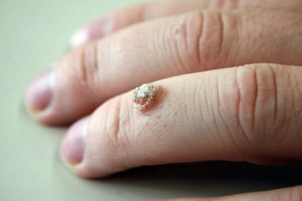
Mastocytosis: what is it, causes, symptoms, treatment, prognosis
Content
- What is mastocytosis?
- Types of mastocytosis
- Signs and symptoms
- Causes and risk factors
- Affected populations
- Symptomatic disorders
- Diagnostics
- Standard treatments
- Forecast
What is mastocytosis?
Mastocytosis is a rare disorder characterized by abnormal accumulation of mast cells in the skin, bone marrow, and internal organs (liver, spleen, gastrointestinal tract, and lymph nodes). Cases beginning in adulthood tend to be chronic and involve the bone marrow in addition to the skin, whereas in childhood this condition is often marked by cutaneous manifestations without the involvement of internal organs and can often be resolved during sexual intercourse ripening. In most adult patients, mastocytosis tends to be persistent and may progress to a more progressive category in a minority of patients. Mastocytosis can be classified into a specific type depending on the symptoms and overall presentation.
Types of mastocytosis
Mastocytosis can mainly affect the skin (called cutaneous mastocytosis) or other parts of the body (called systemic mastocytosis).
- Cutaneous mastocytosis: Cutaneous mastocytosis most often occurs in children. Sometimes mast cells only accumulate in the skin as a single mass (mastocytomas), usually up to 6 months of age. Most often, mast cells accumulate in many areas of the skin, forming small reddish-brown spots or bumps (the so-called pigment hives). Urticaria pigmentosa rarely develops into systemic mastocytosis in children, but it may occur more frequently in adults.
- Systemic mastocytosis: Systemic mastocytosis most often occurs in adults. Typically, mast cells accumulate in the bone marrow (where blood cells are produced). They often also accumulate in the skin, stomach, intestines, liver, spleen and lymph nodes. Organs can continue to function with little or no disruption. But if too many mast cells accumulate in the bone marrow, then the production of blood cells is significantly reduced, and serious blood diseases such as leukemia can develop. If too many mast cells accumulate in the organs, the organs begin to malfunction. The resulting problems can be life threatening.
Signs and symptoms
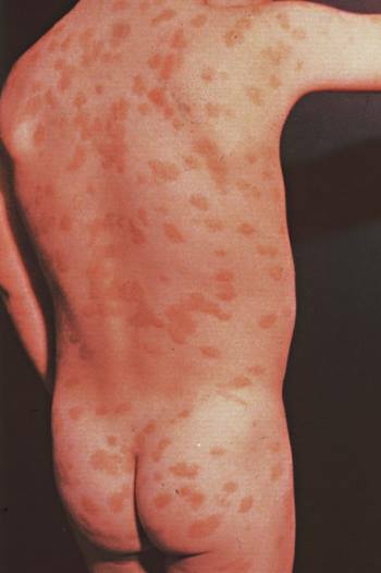
A solitary mastocytoma may not cause symptoms.
Spots and bumps can itch, especially if rubbed or scratched. Itching can be aggravated by:
- temperature changes;
- contact with clothing or other materials;
- the use of certain medicines, including non-steroidal anti-inflammatory drugs (NSAIDs);
- consumption of hot drinks, spicy foods or alcohol;
- physical activity.
If stains are rubbed or scratched, they can develop into hivesand the skin may turn red.
Hyperemia is common.
Peptic ulcer disease can develop due to the fact that too much histamine is produced, stimulating the secretion of excess stomach acid. Peptic ulcer may cause stomach pain. Nausea, vomiting and chronic diarrhea. The abdomen can become enlarged if liver and spleen function is abnormal, which causes fluid to accumulate in the abdominal cavity.
If the bone marrow is affected, bone pain may occur.
People with mastocytosis may become irritable, depressed, or moody.
Widespread reactions may occur. In systemic mastocytosis, extensive reactions are usually severe. These include anaphylactic and anaphylactoid reactions that cause fainting and a life-threatening drop in blood pressure (shock). Anaphylactoid reactions resemble anaphylaxis, but they are not caused by an allergen.
Systemic mastocytosis can affect the bone marrow; in addition, up to 30% of adults with systemic mastocytosis develop cancer, especially myeloid leukemia. Life expectancy in these patients may be short.
Causes and risk factors
Genetic changes (mutations) leading to over-activation of the mast cell growth factor (KIT) receptor have been detected in abnormal mast cells in almost all adults with mastocytosis and in about 80% of children with skin lesions. The most common c-kit mutation in mastocytosis is D816V, and it is believed to cause abnormal proliferation and accumulation of mast cells in tissues. Approximately> 90% of adults and 40% of children are also affected by this mutation, while another 40% of children have mutations in other areas of the KIT. The predictive value of the type of mutation in childhood disease has not yet been determined. Mutations are somatic in nature and therefore are not passed on to the next generation in most patients.
Release of mast cell mediators such as histamine, heparin, chemokines, cytokines, leukotrienes and prostaglandin D2, among other cell mediators, lead to symptomatic episodes. Histamine is a naturally occurring chemical that is released during allergic reactionitchy shortness of breath, dilation of blood vessels and hypersecretion of gastric acid.
Affected populations
Mastocytosis affects men and women in equal numbers. The disorder can begin in childhood or adulthood. The disease in childhood most often manifests itself during the first 2 years of life.
Symptomatic disorders
Symptoms of the following disorders may be similar to those of mastocytosis. Comparison can be useful for differential diagnosis:
- Inflammatory bowel disease (IBD) is associated with an abnormal immune response to naturally occurring bacteria in the gastrointestinal tract. Patients may experience weight loss, abdominal cramps and pain, nausea and vomiting, fatigue, and irregular bowel movements. Diagnosis of inflammatory bowel disease often based on symptoms, which may include a rapid heartbeat (tachycardia), a decrease in the number of erythrocytes (anemia), which leads to fatigue, dehydration and fever. There are various tests and imaging studies that can be performed to confirm the diagnosis and may include: a complete analysis blood tests, serologic tests, stool tests, nutritional assessments, colonoscopy, abdominal ultrasound and other gastrointestinal tests visualization.
- Irritable bowel syndrome (IBS) is a gastrointestinal disorder associated with abdominal discomfort, altered bowel structures and, in some cases, inflammation of the gastrointestinal tract. IBS patients may experience heartburn, nausea and vomiting, clear or white mucus, abdominal pain, and constipation or diarrhea. Patients may also experience bloating, which can be confirmed by computed tomography of the abdominal cavity. There are specific criteria for diagnosing IBS based on the frequency and duration of the disorder's symptoms. The criteria for diagnosing IBS require patients to have recurrent abdominal pain or discomfort for at least three days per month in the past three months associated with two or more of the following conditions: relief from bowel movements; onset associated with a change in stool frequency; and / or onset associated with a change in the shape or appearance of the stool. To diagnose IBS, physical examinations, laboratory tests, and x-rays may be done to rule out any other cause.
- Intestinal malabsorption includes any condition associated with abnormalities during digestion and / or absorption of food nutrients. Patients may experience diarrhea and weight loss; However, the more common symptoms are often based on a specific cause. Various tests can be performed to identify the cause and may include blood, electrolyte and chemical group tests, and serologic testing. Treatment for malabsorption consists of correcting any nutritional deficiencies as well as treating the causative condition.
- Myeloproliferative diseases are a group of disorders associated with the proliferation of one or more separate cell lines. Patients may experience fatigue, weight loss, abdominal discomfort, easy bruising or bleeding, infections, and other symptoms. The specific diagnosis may be based on laboratory tests (i.e., total blood counts, leukocyte alkaline phosphatase readings, analysis polymerase chain reaction, serum uric acid levels, red blood cell mass) and bone marrow biopsies that would reflect changes in cell count blood. Treatment for myeloproliferative disease depends on the specific cause. Patients with chronic myelogenous leukemia can be treated with a number of chemotherapeutic agents. In comparison, treatment is directed towards supportive care for patients with true polycythemia, essential thrombocythemia and myelofibrosis.
- Urticarial rash (hives) Is a skin condition associated with the appearance of red, raised patches on the skin that can itch and irritate when touched. Hives are often isolated and unrelated to other systemic symptoms or signs. Urticaria is often self-limited and transient. Skin lesions can last from 20 minutes to three hours and then disappear within 24 to 48 hours. New-onset urticaria is often associated with an identifiable cause, such as direct contact, and can be identified from the patient's history. Hives can often be confused with others skin diseases. However, a diagnosis can be made based on the characteristic appearance of urticaria (i.e., itchy red skin lesions). Treatment for hives often includes the use of antihistamines.
- Endocrine tumorssuch as carcinoid, pheocromocytoma, and medullary thyroid cancer can cause flushing. Hot flashes after menopause are usually short-lived and associated with sweating.
- Monoclonal mast cell activation syndrome. Have these patients show symptoms of mast cell activation, including recurrent anaphylaxis, but no evidence of cutaneous mastocytosis. Their bone marrow biopsy shows 1 or 2 minor criteria for systemic mastocytosis, but does not meet the full criteria of the World Health Organization (WHO).
- Idiopathic mast cell activation syndrome. Patients with this disorder have episodic symptoms of systemic mast cell activation associated with elevated mast cell mediators such as tryptase and metabolites histamine or prostaglandin in the urine, responding favorably to treatment with drugs that block the mast cell mediator, and have no diagnostic signs of cutaneous or systemic mastocytosis. Other disorders with similar symptoms, such as allergic diseases, must be ruled out before this diagnosis is considered.
Diagnostics
Diagnostics include:
- medical examination;
- biopsy;
- blood tests to rule out other diseases.
Doctors suspect mastocytosis based on symptoms, especially if there are spots that turn into hives and redness when scratched.
A biopsy can confirm the diagnosis of mastocytosis. Typically, a sample of skin tissue is taken and examined under a microscope for mast cells. Sometimes a sample is taken from the bone marrow.
If the diagnosis is unclear, doctors may do the following:
- Blood and urine tests to measure the level of substances associated with mast cells. A high level of these substances confirms the diagnosis of systemic mastocytosis.
- Blood and urine tests to rule out conditions that cause similar symptoms.
- Bone scan.
- Tests for a genetic mutation present in many people with mastocytosis.
- Biopsy (using an endoscope) to determine if there is an abnormally high number of mast cells in the digestive tract.
Standard treatments
Treatment includes:
- drugs to reduce the severity of symptoms;
- for aggressive systemic mastocytosis, other drugs, such as interferon, or surgery, such as splenectomy.
Single mastocytoma can disappear spontaneously.
Itching caused by cutaneous mastocytosis, can be treated with antihistamines. No other treatment is required for children. If adults develop itching and rashes, psoralen (a drug that makes the skin look more sensitive to ultraviolet radiation), and ultraviolet light or corticosteroid creams.
Systemic mastocytosis cannot be completely cured, but symptoms can be suppressed with H1 and H2 blockers. H1 blockers (commonly called antihistamines) can relieve itching. H2 blockers reduce the production of stomach acid and thus relieve the symptoms of peptic ulcers and help heal ulcers. Ingestion of cromolyn can relieve digestive problems and bone pain. Aspirin relieves flushing, but can worsen other symptoms. Children are not given aspirin due to the risk of Reye's syndrome.
If systemic mastocytosis manifests itself in an aggressive form, then subcutaneous injections of interferon-alpha once a week can reduce the effect of the disease on the bone marrow. Ingestion may also help corticosteroids (eg, prednisone), but only for a short period of time. When taken by mouth for more than 3-4 weeks, corticosteroids can cause many side effects, sometimes serious.
If there is a large accumulation of mast cells in the spleen, it may be necessary to remove the spleen.
If leukemia develops, chemotherapy drugs (such as daunomycin, etoposide, and mercaptopurine) may help.
People with systemic mastocytosis should always carry a pen with epinephrine for surgical treatment in the event of an anaphylactic or anaphylactoid reaction.
Forecast
The prognosis depends on the age of onset. Most patients with urticaria pigmentosa show onset before 2 years of age, which is associated with an excellent prognosis, often with resolution at puberty. The number of lesions decreases by about 10% per year. However, acute extensive degranulation can rarely cause life-threatening episodes of shock.
The appearance of cutaneous mastocytosis after 10 years of age portends a worse prognosis, since the disease with a late onset tends to be resistant, is more commonly associated with systemic disease and carries a higher risk of malignant transformation.