Meningioma: what is it, causes, symptoms, treatment, prognosis
Content
- What is a meningioma?
- Signs and symptoms
- Causes and risk factors
- Affected populations
- Related disorders
- Diagnostics
- Standard treatments
- Forecast
What is a meningioma?
Meningioma Is a tumor growing from the cells of the arachnoid meninges, namely the arachnoid endothelium, the tissue that covers the brain and spinal cord. Excluding metastases to the brain from other organs, meningiomas are the most common intracranial tumor and the most common primary brain tumor. They are more common in women and the elderly, and develop in about 8 out of 100,000 people per year. Compression of the brain and / or spinal cord by a meningioma can lead to symptoms such as headaches, seizures, loss of vision, mental changes, and weakness. These tumors are mainly treated with surgical removal (resection), which can be combined with radiation therapy, especially if the location of the tumor is such that it cannot be completely removed.
Introduction.
The term meningioma was coined by Dr. Harvey Cushing in 1922. Since then, the understanding of meningioma has improved significantly and several subtypes have been identified. As of 2019 brain tumors, including meningioma, are classified based on the criteria of the 2016 World Health Organization classification system. Meningiomas are divided into three main categories based on their degree. Tumor grade is determined by microscopic evaluation and refers to the degree of malignancy of the cells of which it is composed: low-grade tumors multiply more slowly and look like normal cells, while high-grade tumors are more aggressive, multiply faster, and do not appear like the cells they are from. happen. According to the 2016 WHO classification, otherwise a grade I meningioma would be classified as grade II in the presence of tumor invasion of the brain. Most meningiomas (80-85%) are benign (grade I), 15 to 18% are atypical (grade II), and 1 to 3% are malignant (grade III). High-grade tumors can arise on their own or as a result of malignant progression of lower-grade tumors.
Signs and symptoms

Meningioma can occur at any age, but it usually occurs in the elderly; the median age at the time of diagnosis is 65 years (meaning that half of the people diagnosed will be older than this age and the other half will be lower). Potential symptoms of meningioma are variable and depend on the location of the tumor, as well as on the compromising areas of the brain and spine.
Parasagittal meningioma can lead to symptoms such as headaches, seizures (epilepsy) and weakness of the limbs. Pressure within the skull (intracranial pressure) can be increased and cause swelling of the optic nerve and blurred vision. Other visual defects can develop as well. Compression on the frontal lobe can lead to personality changes, decreased cognition, and a lack of emotion and interest (apathy). Involvement of the cerebellum can lead to gait imbalances (ataxia), decreased coordination (dysmetria), and involuntary eye movements (nystagmus).
Hearing loss can occur if the tumor is located at the junction between the cerebellum and an area of the brain stem known as the bridge. A tumor located in the opening of the skull where the spinal cord connects to the brain stem (large opening) can also cause weakness in the arms and legs. Compression by meningiomas can also impair the removal of cerebrospinal fluid and lead to accumulation of cerebrospinal fluid in the brain, a condition known as hydrocephalus.
About 10% of meningiomas occur in the spine (spinal meningiomas), most often in the thoracic region. Symptoms associated with spinal cord injury include difficulty walking, weakness and numbness in the legs, pain, and difficulty urinating and / or defecating.
Causes and risk factors
As the name suggests, meningioma comes from meninges, which are membranes that cover the brain and spinal cord. More specifically, most meningiomas stem from a type of cell in the meninges known as the arachnoid membrane cells, which are involved in the resorption of cerebrospinal fluid.
Although the exact causes of meningioma in some people are in most cases unknown, it is believed that several molecular mechanisms are involved in the development of meningioma. In meningioma cells, several genes are often altered (mutated), one of the most important of which is NF2. This gene produces merlin, a tumor suppressor protein that reduces (inhibits) the growth of cells that are in contact with each other. It has also been shown that meningioma cells express the protein survivin, which is an inhibitor of programmed cell death (apoptosis). Growth factors and growth factor receptors such as platelet growth factor and HER2also play a role in the development of some meningiomas. Overexpression tumors HER2appears to be associated with a higher relapse rate.
Meningioma has other features that are common to many types of tumors: they tend to generate numerous blood vessels, which will increase the flow of nutrients that can help growth. Vascular endothelial growth factor (VEGF) is involved in the development of new blood vessels (angiogenesis) and is often overexpressed in numerous tumors, including meningioma. A characteristic feature of malignant meningioma is an increased ability to lengthen telomeres due to mutations in the gene TERT. Telomeres are located at the end of the chromosome and shorten with each cell division until they are too short for cell division. They can be lengthened by the enzyme telomerase, which gives an increased replication potential for cells and promotes tumor growth. TERT the gene encodes a subunit of the telomerase enzyme.
Some meningiomas carry progesterone, estrogen and androgen receptors, indicating that hormones could potentially be involved in the growth of some meningiomas. Other factors that indicate the role of sex hormones in meningioma is the fact that they are more common in women and that associated symptoms may worsen during periods of excess progesterone, such as pregnancy. The role of sex hormones in the development and growth of meningioma, however, is not well understood.
Affected populations
Meningioma accounts for about a quarter to a third of primary intracranial tumors (excluding metastases from other tumors) and develops in about 8 out of 100,000 people per year. As of 2010, 170,000 people have been diagnosed with meningioma in the United States alone. There is no data for Russia. However, this is likely an underestimate, as US autopsy data show that up to 2.8% of people have a meningioma. However, the vast majority of these tumors are too small to cause symptoms and will never grow large enough to be clinically significant.
Meningioma is most common in older adults, with a median age at diagnosis of 65 (this means that half of the people diagnosed will be older than this age, and the other half will be lower). These tumors are rare in children. It is noteworthy that they arise in connection with syndromes such as type II neurofibromatosis. (See Related Disorders for more information.) They occur 2-3 times more often in adult women, but are equally common in boys and girls, as well as in the case of malignant meningioma. Spinal meningioma occurs in a 9: 1 female: male ratio. Meningioma is also slightly more common in blacks. Another well-established risk factor is prior exposure to ionizing radiation, for example, in people who have previously received radiation therapy to the head. Radiation-associated meningioma is more often atypical or malignant and multiple. In most cases, they occur at least 20 years after exposure.
Related disorders
Disorders associated with meningioma can be divided into two categories: syndromes with multiple symptoms and signs, one of which is increased risk of meningioma, and diseases that are not meningiomas, but which may show similar meningiomas in patients symptoms.
— Syndromes associated with the development of meningioma.
- Type 2 neurofibromatosis (NF2) is a rare genetic disorder primarily characterized by benign tumors originating from Schwann cells (schwannoglioma), which are cells that form an insulating myelin sheath around nerves outside the brain and spinal cord (peripheral nerve system). In NF2, schwannoma usually forms around both vestibulochular nerves, which transmit balance and sound impulses from the inner ears to the brain. These tumors are called vestibular schwannomas or acoustic neuromas. People with NF2 are also at increased risk of developing cataracts and some tumors of the brain and spinal cord, including meningiomas.
- Schwannomatosis - a disorder characterized by the presence of multiple schwannomas, almost always without vestibular schwannomas, which are diagnosed in NF2. Patients with schwannomatosis may develop benign tumors in the brain (including meningioma) and along the spinal or peripheral nerves.
- Meningioma is also more common in people with cancer predisposition syndromes. These syndromes are multiple and include Gorlin syndrome, Cowden syndrome, Li-Fraumeni syndrome, and von Hippel-Lindau syndrome.
- Diseases with symptoms similar to those of a meningioma.
Other types of brain tumors have symptoms similar to meningiomas. They are classified according to their origin. Examples include gliomas, medulloblastomas and brain metastases, which are more common than tumors arising in the brain (primary tumors brain), and come from cancers in other parts of the body, including the lungs, breasts, and gastrointestinal tract.
Intracranial abscesses can also lead to neurological symptoms similar to those present in meningiomas. Causes brain abscesses are plural and include bacterial meningitis, fungal and parasitic infections (cryptococcosis and cysticercosis, respectively), septic cerebral embolism and brain inflammation (encephalitis).
Diagnostics
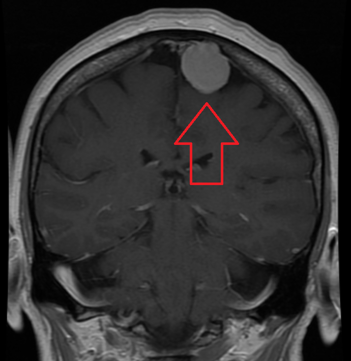
The diagnosis of meningioma is mainly based on the patient's medical history and physical examination, medical imaging, and analysis of the cells that make up the tumor. A patient who has signs and symptoms suggestive of central nervous system involvement, such as recent epileptic seizures or neurologic deficiency, usually undergoing medical imaging of the brain and / or spine using computed tomography (CT) or magnetic resonance imaging (MRI).
MRI is the preferred imaging technique for the primary assessment of brain tumors. Although meningiomas can be suspected by medical imaging due to their characteristic location, analysis of tumor cells provides the most accurate diagnosis. Meningioma cells can be obtained by removing part of the tumor with a biopsy.
However, meningiomas are usually removed surgically without prior biopsy, as this provides a therapeutic benefit in addition to a diagnostic benefit. Once the tumor cells are obtained, they can be examined under a microscope by a pathologist who will confirm the diagnosis. A laboratory technique called immunohistochemistry can be used to facilitate identification of meningioma cells.
Standard treatments
Standard treatment for meningioma - complete removal (resection) of the tumor. This procedure is performed by a neurosurgeon; The doctor specializes in the surgical treatment of diseases of the nervous system. To gain access to the brain for tumor resection, a portion of the skull is removed (craniotomy) and returned to the site after surgery. If the tumor has a large blood supply, which may complicate surgery, it can be used before surgery a technique called angiography to block (embolize) the blood vessels that feed the tumor in order to relieve it resection.
Various surgical techniques can also be used to treat spinal meningioma. Often, one operation is sufficient. However, if the localization of the tumor is such that it cannot be completely removed due to its proximity to important structures, it can be used radiation therapy alone or in combination with surgery. Radiation Oncologists - doctors who specialize in the use of radiation therapy for the treatment of tumors and cancers. Radiation therapy involves the use of high-energy X-rays or other types or energy to destroy tumor cells.
For meningiomas, external beam radiation therapy is most commonly used. Many devices can be used for external beam radiation therapy, but the goal is the same in all cases: to target the tumor and expose it to multi-beam radiation to destroy it while avoiding damage to healthy brain tissue as much as possible Maybe. Because of the high risk of recurrence, radiation therapy is often used deliberately after surgery for many atypical meningomas and regularly for all malignant meningomas, even if the tumor was completely removed. Radiation therapy can be used alone if the tumor cannot be surgically removed.
Chemotherapy is not usually used to treat meningioma. However, hydroxyurea, an old chemotherapy agent, is sometimes used when surgery and radiation therapy have been depleted. However, it has shown a limited therapeutic response. Other medications that can be used to treat meningioma include prescribing blood thinners (anticoagulants) such as heparin to reducing the risk of a blood clot (thrombosis) after surgery or anticonvulsants if the affected person develops convulsions.
After treatment, patients may require rehabilitation to restore function affected by the tumor or its treatment. Rehabilitation teams include many health professionals, including doctors, physiotherapists, occupational therapists and nurses.
Forecast
Since most meningiomas are benign, the survival rates of sick people are relatively high: more than 80% of patients survive for more than 5 years, about 75% survive for more than 10 years, and about 70% survive for more 15 years. Older age, male gender, poor baseline health, and high-grade tumors are associated with lower life expectancy.
Lower-grade meningiomas that are completely removed by surgery usually do not recur. However, some tumors cannot be completely removed due to their location and proximity to important structures. The rule of thumb for recurrences of incompletely resected meningiomas is that the recurrence rates are 30%, 60%, and 90% at 5, 10, and 15 years, respectively. Resected higher-grade tumors may recur even with apparent complete resection, most often within 2 or 3 years. Tumors that have already recurred are more likely to recur, usually within a shorter period of time. Metastases to the spine and other organs are rare in meningiomas and are most commonly associated with grade III tumors.