Fibrous dysplasia: what is it, causes, symptoms, treatment, prognosis
Content
- What is fibrous dysplasia?
- Signs and symptoms
- Causes
- Diagnostics
- Standard treatments
- Forecast
What is fibrous dysplasia?
Fibrous dysplasia (FD, Jaffe-Liechtenstein disease) is a rare bone disorder. The bone affected by this disease is replaced by abnormal scar (fibrous) connective tissue. This abnormal fibrous tissue weakens bone, making it abnormally fragile and prone to fracture. Pain may occur in the affected areas of the body. As the patient grows older and grows, the affected bone may become deformed (dysplastic).
PD can affect only one single bone (monostatic disease), or the disorder can be widespread, affecting multiple bones throughout the body (polyostatic disease). The severity of the disorder can vary greatly from one person to the next. Any part of the skeleton can be affected, but most often the long bones of the legs, bones of the face and skull (craniofacial region), and ribs are affected.
Fibrous dysplasia is usually diagnosed in children or adolescents, but mild cases may not be diagnosed until adulthood. In some cases, fibrous dysplasia may not require treatment; in others, certain medications and surgical procedures may be recommended.
Signs and symptoms
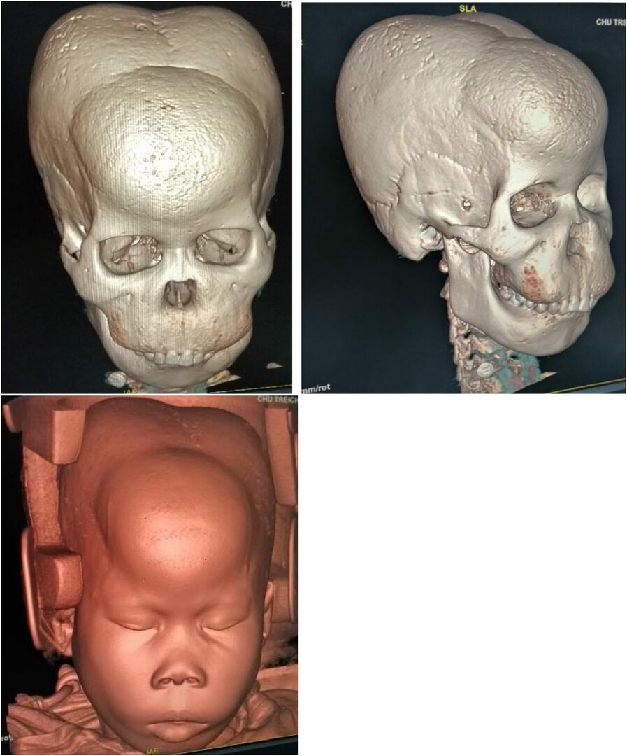
Fibrous dysplasia often has no pronounced symptoms. The patient may not be aware of the disease for a long time, since painful sensations and inconveniences do not arise until a certain level of growth of the disease. The disease can affect one bone or an entire skeletal system. The peculiarity of damage caused by fibrous dysplasia is determined by the amount of cellular and fibrous tissue dotted with blood vessels, and scattered bone formations, around which there is bone-like tissue, in turn, surrounded by cells. Two types of growths in the bones can be distinguished: focal and diffuse.
In the first, focal lesion, the transformed part is detached from the surrounding tissues and easily moves away from the adjacent bone. The tissue that has undergone transformation becomes dense, but does not lose its elasticity, turns pale, bone inclusions are clearly visible in it. With the second, diffuse bone lesion, it is impossible to determine the size of the damaged tissue, due to the fact that the adjacent bone becomes porous and soft, the cortical layer becomes thinner. The porous tissue of the bone is replaced by a dense, pale yellow substance. In both types, it is possible to detect single cysts.
Read also:Heel spur, causes, symptoms and home treatment
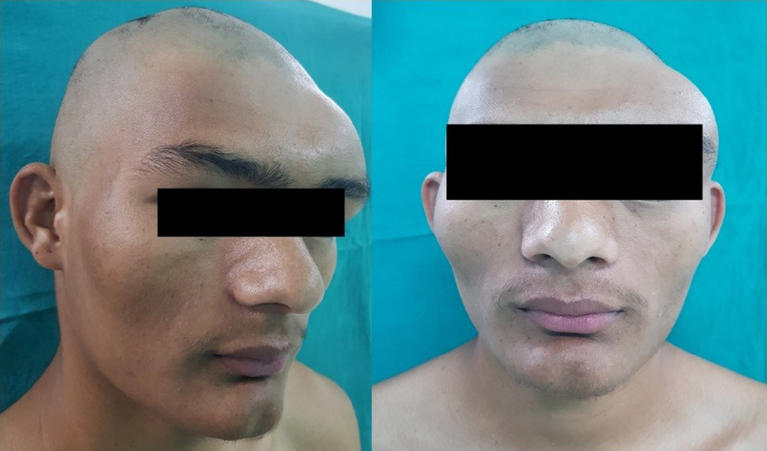
Clinical manifestations of a single bone lesion are subtle, do not have clear symptoms. The infected area gradually becomes thicker, while no pain occurs. The disease becomes noticeable after about 2 years from the onset of the inflammatory process. Asymmetry often appears. When the disease manifests itself on several bones at the same time, the symptoms remain the same, but the area of the lesion increases significantly. Frequent fractures are one of the most common signs, and skin pigmentation is also possible.
Causes
The main reason for FD is not fully understood. Researchers believe the disorder is caused by a mutation in a gene called GNAS1. This gene mutation occurs after fertilization of the embryo (somatic mutation) and therefore is not inherited and affected people will not pass the mutation on to their children. Affected people have some cells with a normal copy of this gene and some cells with an abnormal gene (mosaic pattern). The variability in the symptoms of fibrous dysplasia is partly due to the ratio of healthy cells to abnormal cells. Researchers don't know why these somatic mutations occur; they develop randomly for unknown reasons (from time to time).
Gene GNAS1 located on the long arm (q) of chromosomes 20 (20q13.2). Chromosomes, which are present in the nucleus of human cells, carry the genetic information of each person. The cells of the human body usually have 46 chromosomes. Pairs of human chromosomes are numbered 1 through 22, and the sex chromosomes are labeled X and Y. Men have one X and one Y chromosome, while women have two X chromosomes. Each chromosome has a short arm, labeled "p", and a long arm, labeled "q". Chromosomes are further subdivided into many numbered bands. For example, "chromosome 20q13.2" refers to band 13.2 on the long arm of chromosome 20. The numbered stripes indicate the location of the thousands of genes present on each chromosome.
Gene GNAS1 creates (encodes) a protein known as the G protein. In PD, gene mutation GNAS1 leads to an overproduction of this G-protein. This in turn leads to an overproduction of a molecule known as cyclic adenosine monophosphate (cAMP), which is involved in the alteration (differentiation) of osteoblasts in bone. Osteoblasts are bone-forming cells that form new bone. The human skeleton is a living tissue that is constantly changing (reconstructed). PD is thought to include an increase in bone turnover. Bone turnover is a normal process in which bone is gradually destroyed (bone resorption) and then restored. Bone turnover includes osteoblasts and cells that control bone resorption (called osteoclasts). The interaction between osteoclasts and osteoblasts determines how bones are transformed. Interaction is a complex process that includes many factors. It is believed that maldifferentiation of osteoblasts due to gene mutation GNAS1 promotes the development of PD. Osteoclast bone-removing activity likely allows skeletal progenitor cells, including immature osteoblasts and fibrous tissue, to have more room for growth and reproduction.
Read also:Hartnup's disease
When other cells, such as endocrine or skin cells, are involved in the process in addition to osteoblasts, McCune-Albright-Braitsev syndrome develops.
Diagnostics
FD can be diagnosed using an X-ray machine and histological analysis. With histological examination, the continuity of transformations in the tissues is noted during the period of puberty, which often occurs earlier in infected people. When examining with an X-ray machine, lesions are revealed, filled with bone tissue of different density. In some cases, skin pigmentation over the affected area becomes noticeable.
Standard treatments
In the overwhelming majority of cases, if the deviations are not pronounced, and the diagnosis is carried out at an early stage of fibrous dysplasia, the treatment gives positive results. For some patients, standard therapy will be sufficient, while for the treatment of others, curettage and graft repair should be used. Failures are due to resorption of the graft and recurrence of initially diagnosed symptoms.
Fibrous dysplasia of the femur in neglected condition leads to a change called "shepherd's stick". With such a violation, it is necessary to carry out a local operation to remove the deformed area and replace it with a suitable graft. In the absence of pathologies and relapses, treatment is successful.
Fibrous dysplasia of the skull in a neglected state turns into a type called "lion's face". With such a localization of the disease, the bones seem to swell. If such fibrous dysplasia is found in children, then facilitated surgery is indicated for elimination of further deformation of the facial bones, while taking into account their subsequent growth and development as they grow the patient.
PD of the tibia is externally manifested by lameness and gait disturbances; shortening of the limb, in which the affected bone tissue is located, is rarely noticed. Treatment also requires surgery, plastic surgery and follow-up. After the operation, patients are forced to use crutches and orthopedic devices until complete or partial recovery.
Read also:The bone on the big toe: causes and treatment (methods, recipes of traditional medicine)
The degree, duration and intensity of therapy can only be determined by a doctor specializing in the treatment of this disease. The method of treatment is selected for each patient exclusively individually. For children, due to the fragility and insufficient development of the skeletal system of the child's body, therapy is first of all used, and only if it is ineffective, an operation is prescribed. An adult patient is diagnosed with the severity of the course and the neglect of the development of the disease. In most of the known examples, the operation is carried out immediately. If one bone is damaged, it is removed, replacing it with a graft. With multiple lesions, a longer and more serious treatment with the use of high-tech devices is required.
In general, after undergoing therapy, an operation, and the introduction of a graft, the patient becomes much better. But we cannot stop there. Constant supervision by a specialist is necessary, which will reduce the likelihood of relapse, up to the patient's complete recovery.
Forecast
People with milder forms of PD often live normal, otherwise healthy lives. The prognosis is as varied as the disease itself, and is based on damage to bones, damage to other structures such as nerves, and the presence of fractures.
Fibrous dysplasia can affect many bones in the body, but once it has formed in the skeleton, the disease does not spread.