Acanthamebic keratitis: what is it, symptoms, treatment, prognosis, complications
Content
- What is acanthamoebic keratitis?
- Symptoms and Signs
- Pathogenesis
- Life cycle
- Epidemiology
- Diagnostics
- Standard treatments
- Forecast
- Complications
What is acanthamoebic keratitis?
Acanthamebic keratitis (AK) is a rare vision-threatening disease that occurs primarily in contact lens wearers. The disease is caused by an infection of the protozoa acanthamoeba (acanthamoeba) and is commonly mistakenly identified as one of the most common fungal or viral forms keratitis, especially ocular herpes (herpetic keratitis).
Accidental corneal injury, which is more common in developing countries, and contact lens wear are major risk factors for AK.
Treatment methods are most effective in the early stages, so timely diagnosis is critical to stop disease progression. In later stages, patients often face corneal transplant as the only way to restore visual acuity; thus, acanthamoebic keratitis should be of major concern in contact lens wearers and those who have recently suffered from surgical or accidental corneal injury.
Prevention is the best weapon against AK, as in most cases the disorder can be directly related to poor contact lens hygiene or direct eye contact with contaminated water.
Symptoms and Signs
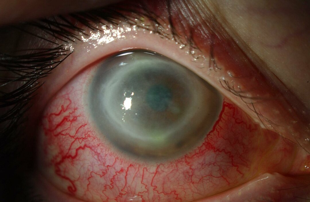
Patients with AK usually complain of redness, pain, decreased visual acuity and sensitivity to light (photophobia). The most prominent clinical feature of acanthamoebic keratitis is a pronounced annular stromal infiltrate, which is believed to be composed of inflammatory cells. As the disease progresses, signs of scleritis, corneal inflammation and conjunctival hyperemia begin to appear.
If left untreated, acanthamoeba can spread back to the retina and cause severe chorioretinitis, and in these advanced stages, patients often experience reduced visual acuity or complete blindness. Late diagnosis, severe penetration, and deep stromal infection are all associated with poor visual outcomes. Patients who develop stromal keratitis are also more likely to have poor vision after treatment than patients with more superficial epithelial infection.
Pathogenesis
In Russia and other developed countries keratitiscaused by acanthamoebais almost always associated with the use of soft contact lenses. Acanthamoeba spp. most often gets into the eyes through contact lenses that have been exposed to the parasite as a result of using a solution with contaminated lenses, household saline solution or tap water, or from wearing contact lenses while bathing or swimming. However, the parasite can also get into the eyes through contact with soil or vegetation or grass.
Read also:Allergic conjunctivitis
In fact, the first reported case of AK was caused by an eye injury. Once on a contact lens acanthamoeba able to survive in the space between the contact lens and the surface of the eye. Soft contact lenses adhere better to the surface of the cornea than hard contact lenses, allowing the body to acanthamoeba bind to mannosylated glycoproteins on the surface of the cornea. The expression of these proteins on the surface of the cornea is increased with contact lenses. This increase in glycoprotein content, along with microtrauma to the epithelial surface of the cornea due to the use of contact lenses, increases the risk of infection.
Once the body has gained access to the surface of the eye, it can penetrate the Bowman's membrane (membrane). In some cases, the infection can cluster around the corneal nerves, causing radial deposits (radial keratoneuritis) and severe pain. These features are also seen in viral and bacterial keratitis and can be misleading. The parasite is also able to penetrate deeper into the cornea; with the help of metalloproteases, acanthamoebas are able to penetrate deep into the stroma of the cornea. As the disease progresses, the parasite can penetrate the cornea, and very rarely causes an infection inside the eye (endophthalmitis) due to a strong reaction of neutrophils in the anterior chamber of the eye.
While the vast majority of AK cases occur in contact lens wearers, many cases of acanthamoeba in those who do not wear contact lenses, especially outside the developed world. Non-contact lens wearers are at greatest risk of infection acanthamoeba Are injuries and exposure to contaminated water. Other contributing factors include contaminated water supply in the home and low socioeconomic status. The infection is also more common in tropical or subtropical climates.
In addition to the route of inoculation into the eye and external risk factors, host factors may also play a significant role in the development of acanthamoebic keratitis. In fact, studies of contact lens wearers in the United Kingdom, Japan and New Zealand have shown that 400 to 800 out of 10,000 asymptomatic contact lens wearers had lens containers, infected acanthamoeba spp. However, the incidence of acanthamoebic keratitis among these patients was only 0.01–1.49 per 10,000 contact lens wearers. Although the exact host factors have not been fully described, it is likely that defects in corneal epithelium, tear film composition, eye surface pH and anti-cantal IgA antibodies in the tear film play a role in the development of acanthamoebic keratitis.
Read also:Amblyopia in children
Life cycle
Species within the genus acanthamoeba, as a rule, are free-living trophozoites. These trophozoites are relatively widespread and can live, but are not limited to, in tap water, freshwater lakes, rivers, and soil. In addition to the trophozoite stage, the body can also form double-walled cysts, which can also be present in the environment and are very difficult to cure with medical treatment. Both of these stages usually do not originate and are reproduced by means of binary fission.
Epidemiology
A study from Austria reported a total of 154 cases of acanthamoebic keratitis over a 20-year period. The age of patients with AK ranged from 8 to 82 years, 58% of people were women. The data showed that 89% of infected patients wore contact lenses, almost all cases occurred in only one eye, and 19% required corneal transplantation.
Diagnostics
Correct early diagnosis is important before acanthamoeba will penetrate deeper into the cornea and cause irreversible tissue damage. Unfortunately, at the first diagnosis, the disease is more often diagnosed as herpetic keratitis, and not acanthamoebic. This misidentification can lead to the use of antiviral drugs, which will only exacerbate the patient's symptoms. At the time of diagnosis, acanthamoba is often viewed as a cause of infection when corneal ulcers do not respond to treatment with antiviral and antibacterial drugs. This delay in making a correct diagnosis allows the parasite to move further into the eye.
The first step to correct diagnosis is an accurate examination with a slit lamp. Slit lamp may show signs of acanthamoeba either through its direct presence or through an associated immune response. The absence of bulbous dendrites characteristic of herpes patients is a good indicator that the patient may have AK. Early and accurate examination with a slit lamp is important for patients with corneal ulcers as the stage disease at the time of diagnosis AK is a strong predictor of final visual result.
If possible, direct in vivo diagnosis can be performed using an advanced tandem scanning confocal microscope and a Heidelberg retinal tomograph. Polymerase chain reaction (PCR) can also be used as a diagnostic tool when too few cells are available for accurate visual identification. If research in vivo and PCR are not feasible, cultivation of corneal scrapings can be a valuable diagnostic tool acanthamoeba and differentiating it from ocular herpes.
Read also:Macular degeneration of the retina (macular degeneration)
Standard treatments
Treatment is usually done with antiseptic drops, including polyaminopropyl biguanide, chlorhexidine, propamidine isethionate (Brolen), or hexamidine, which have anti-amebic properties. Typically, the patient needs to take these eye drops every hour for the first few days (including at night), reducing to 2 hours a day, and then less frequently as treatment progresses. It can be quite difficult to take the eye drops at night for the first few days, but it is very important to try to stick to the regimen indicated by your doctor.
In addition to anti-aesthetic eye drops, anti-inflammatory or pain relieving medications are prescribed to relieve pain. In the early stages of the infection, dilating drops may be prescribed to stop painful spasms in the colored part of the eye, the iris. About 10% of acanthamoeba infections have dual pathology, which means that another infection is present, usually bacterial. In this case, antibiotics and other drops are prescribed. Sometimes antibiotics are also prescribed to protect against bacterial infection when the surface of the eye is damaged in the early stages of the disease. Patients with severe inflammation or scleritis (acute autoimmune inflammation of the deep layers of the sclera of the eye) sometimes prescribe steroid eye drops, although not every patient requires them, and their use requires careful control.
Forecast
The most important prognostic factors for acanthamoebic keratitis are the severity of the disease at the time of admission and the time required to initiate effective therapy. Most patients have a poor prognosis due to a significant delay in diagnosis, which leads to corneal scarring; however, if treatment is started within three weeks of symptom onset, patients usually have good visual results. The presence of a cataract or lesion outside the cornea indicates a worse prognosis.
Complications
Common complications of acanthamoebic keratitis include glaucoma, iris atrophy, wide anterior synechiae, cataract and persistent endothelial defect. Rarer complications include scleritis, sterile anterior uveitis, chorioretinitis and vasculitis retina. Scleritis occurs in about 10% of acanthamoebic keratitis cases and is considered secondary to an inflammatory response of unknown etiology rather than direct invasion acanthamoeba. Extracorneal inflammation should be controlled with the anti-inflammatory drugs listed above.