Erythema multiforme: what is it, symptoms, causes, treatment, prognosis
Content
- What is erythema multiforme?
- Signs and symptoms
- Stages
- Causes and risk factors
- Epidemiology
- Pathophysiology
- Histopathology
- Diagnostics
- Treatment
- Forecast
- Complications
What is erythema multiforme?
Erythema multiforme (ME) is a hypersensitivity reaction of the skin and mucous membranes with characteristic target-like eruptions, caused by certain antigenic stimuli. The disease is an acute, sometimes recurrent condition of the skin and mucous membranes, manifested by papular, bullous and necrotic lesions. Its causes are varied and numerous, and mild cases are generally favorable.
Most lesions appear after 48 to 72 hours and appear on the limbs. The rash remains localized in one area and heals within 7-21 days. Common triggers include herpes simplex virus, histoplasmosis and Epstein-Barr virus. Recurrences are not uncommon if the trigger is herpes simplex. Although most cases are mild, severe cases can be life-threatening. Mucous membranes can be affected in 2-10% of people. In general, most cases of erythema multiforme are medication-related.
Signs and symptoms
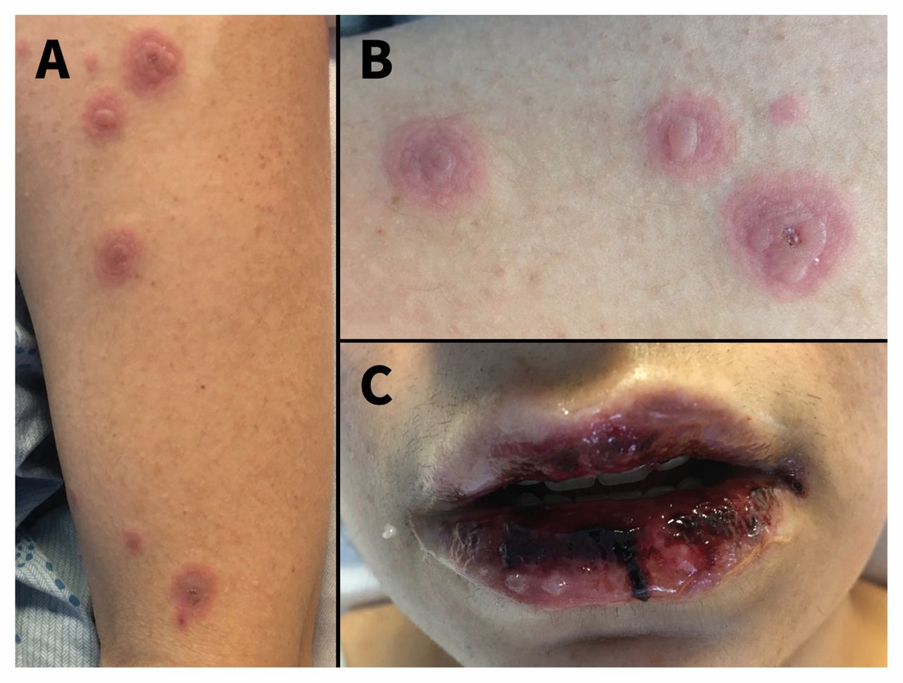
Fever and general anxiety may precede and / or accompany the rash in the early days. Sometimes arthralgia occurs or even edema joints.
The clinically typical ME lesion is target-like lesions, described as rounded lesions that have three concentric circles and a well-defined border. The peripheral ring is erythematous, sometimes microvesicular; the middle zone is often clearer, edematous and palpable, and the center is erythematous, blistered. These different aspects cause different stages of the developing lesion.
The lesions are less than 3 centimeters in size and are mostly acral. They are symmetrical on the palms and the back of the hands, feet, and outstretched surfaces of the limbs. The trunk is often not affected, but the face and ears are often affected. There is no itching, but some patients experience a burning sensation.
Lesions of the mucous membranes are common, mainly in the mouth, but also on the mucous membranes of the genitals and eyes. At first, they are bullous, then quickly turn into painful erosion. Thick hemorrhagic crusts can cover lip lesions, and fibrinous whitish plaque can cover erosions of the mucous membranes of the cheeks, palate, and genitals. These mucosal lesions most often occur concurrently with skin lesions, but may move several days before or after the eruption of the target eruption. While skin lesions are painless, mucosal lesions are often painful.
Read also:Acrocyanosis
Pulmonary symptoms such as coughing and dyspnea (dyspnea). They indicate a respiratory attack, most often associated with an infection causing erythema multiforme (mainly due to Mycoplasma pneumoniae).
With extensive skin lesions, some patients may experience signs of dehydration. Others with mucosal lesions may lose weight due to difficulty eating.
Stages
- Small erythema multiforme: in practice, it affects only the skin with typical lesions of a symmetrical acral arrangement. Mucosal involvement is rare and, when present, is mild and affects a single mucosa, often the oral cavity.
- Large erythema multiforme: skin lesions are more extensive, but do not exceed 10% of the body surface area. Typical targeted eruptions are present. Mucosal involvement is severe and affects at least two different areas of the mucosa; the mucous membrane of the mouth is usually affected.
Causes and risk factors
The etiology of ME is dominated by herpes simplex virus (HSV) types 1 and 2 and Mycoplasma pneumoniaebut many other viral, fungal and bacterial infections are involved. Less commonly and more dubiously, vaccines are accused.
Medicines associated with erythema multiforme include antibiotics such as penicillins, cephalosporins, macrolides, sulfonamides, anti-tuberculoid drugs, antipyretics and a lot others. In some patients, exposure to heavy metals, herbs, topical medications, and poison ivy can cause ME.
Epidemiology
Erythema multiforme occurs worldwide without any ethnicity. The disease occurs at any age, more often in young people. The average age is between 20 and 30 years old, and 20% of cases are in children. ME is more common in men with a sex ratio of 1 to 5. The prevalence is unknown, but appears to be well below 1%. Since the classification is not always clear, cases Stevens-Johnson syndrome were often included in ME studies.
HIV patients taking corticosteroidsimmunocompromised who have undergone bone marrow transplantation and lupusare prone to the development of erythema multiforme.
Pathophysiology
Erythema multiforme is often associated with viral or bacterial infections, especially herpes simplex virus (HSV / HLA). Studies have demonstrated the presence of HSV DNA by polymerase chain reaction in acute or secondary ME lesions. The contributing factors are unknown. HLA-DQ3 has been reported to be associated with postherpetic ME and has been suggested as an additional diagnostic marker. Other groups of human leukocyte antigens have also been reported as markers of recurrent erythema multiforme.
Read also:Dry skin - causes, symptoms, diagnosis and treatment
Damage to epithelial cells occurs through cellular immunity. In the early stages of the disease, there is an influx of macrophages and CD8 T-lymphocytes, which release a wide range of cytokines that mediate inflammation and, as a result, cell death.
When the process is caused by drug hypersensitivity, the earliest pathological sign is keratinocyte necrosis.
Histopathology
Puncture biopsy can be used to confirm the diagnosis of ME. The classic lesion is revealed by the vacuolar interface dermatitis with pronounced infiltration of lymphocytes along the dermoepidermal junction. In addition, dyskeratosis of basal keratinocytes and hydropic changes can be observed. With advanced lesions, epidermal necrosis, subepidermal blisters and vesicles are usually observed. CD 8 T-lymphocytes and macrophages predominate.
Diagnostics
The diagnosis of ME is clinical. In case of doubt, a biopsy of the skin of the lesion may be performed for histological examination with immunofluorescence. He then shows epithelial intercellular edema with necrosis of keratinocytes, responsible for an intra- or sub-epidermal blister covered with necrotic epidermis. Perivascular lymphohistiocytic infiltrate is present in the superficial dermis without necrotic vascular lesions. Direct immunofluorescence is negative. Biological evaluation provides no evidence for a diagnosis of ME. However, it is useful to assess the severity of the disease. Chest X-ray may show interstitial radiologic infiltration (mainly in ME due to Mycoplasma pneumoniae). Also described are renal, hepatic, or hematological lesions that are not systematically observed in milder forms.
Treatment
Acute phase treatment:
- Local treatment is based on the use of antiseptics for bullous lesions, antiseptic mouthwashes and anesthetics. Eye lesions are treated by ophthalmologists. Healing is facilitated by applying petroleum jelly to the lips and ointment with vitamin A to the eyes.
- General treatment is used in cases of general condition and dietary difficulties requiring hospitalization for pain relief and hydration. The question of the place of systemic corticosteroids and intravenous immunoglobulins has been discussed without demonstrating their effectiveness. With extensive lesions, daily monitoring is necessary.
- Etiological treatment should be started when the cause is established (or sometimes probable). Mycoplasma pneumoniae infection justifies treatment with azithromycin for three days without even waiting the results of bacteriological examination, especially if there is a cough or radiological abnormalities in lungs. Some suggest treating herpes with acyclovir or valacyclovir if herpes is suspected, although this has not generated interest.
Prevention of recurrent forms of ME:
- In most cases, it is herpes. Even if evidence has not been supported by samples, long-term treatment with acyclovir or valacyclovir should be suggested.
- Theoretically, more than 5 ME flashes per year are indicated. Treatment with valacyclovir prevents herpes simplex virus outbreaks of ME, but does not appear to have an effect on an ME outbreak if it starts after the onset of the rash.
- If germs are not found, other therapeutic options may be suggested in the long term. drugs such as hydroxychloroquine, dapsone or early fetal systemic corticosteroids.
Read also:Scrofula in adults and children: what is this disease, symptoms and treatment of the disease
Forecast
The prognosis is mainly related to the detached surface of the body. Healing is achieved spontaneously after 2-3 weeks with a small erythema multiforme and after 4-6 weeks with a large one. Mucosal lesions always take longer to heal. Healing of mucocutaneous lesions occurs without scarring, but with frequent dyschromia. Relapses are observed in less than 5% of cases, mainly in the forms caused by herpes infection.
The main long-term risk is the development of synechia with lesions of the mucous membranes. Eye complications can be serious and lead to blindness. At the genital level, synechiae can have functional consequences.
During an acute attack, special vigilance must be exercised to prevent these consequences. The prognosis is only vital in exceptional cases when the treatment is adapted. Two situations deserve special attention: (1) severe mucosal involvement and (2) bacterial superinfection.
Poor prognostic factors include impaired renal function, previous bone marrow transplantation, damage to internal organs, and old age.
Unfortunately, some patients may develop an ongoing form of the disease that does not respond to treatment. It can happen in patients with HSV infection, reactivation VEB, inflammatory bowel disease and latent renal cell carcinoma.
Complications
While mucosal lesions heal completely, skin rashes can lead to scarring. In addition, strictures of the urethra, esophagus, vagina and anus are not uncommon. Urinary retention has been reported. phimosis and hematocolpos as a result of strictures. Eye complications occur in 20% of patients and can lead to uveitis, conjunctivitis, corneal scarring, panophthalmitis and permanent blindness. In case of narrowing of the nasolacrimal duct, an epiphora may occur. Many patients develop dry eye syndrome and corneal scarring.