Retrocecal appendicitis: symptoms, treatment, clinical picture and diagnosis
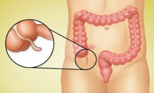
Retrocecal appendicitis is an inflammation of the appendage of the large intestine, non-standardly located along the posterior surface of the intestinal wall, in a retrocecal pocket. It is not anatomically separated, but isolated due to the occurrence of various kinds of pathologies - adhesions, adhesions - retroperitoneal pocket. The very concept of appendicitis is an acute pathology, characterized by inflammation of the appendix of the intestine (an anatomical formation, the length of which ranges from 4 to 10 cm, and the diameter up to 6 mm). The course, manifestation of pathology is similar in symptoms to ordinary food poisoning.
The appendix fits snugly to the posterior surface of the intestinal wall and has a short mesentery. The structure is tubular, originates from the end cecum. Symptoms vary depending on the adhesion of the appendix.
Content
- 1 Types of appendicitis
- 2 Symptoms
- 3 How and what to treat
-
4 Clinic
- 4.1 Clinic for the elderly, children and pregnant women
- 5 Diagnosing appendicitis
Types of appendicitis
- Retroperitoneal appendicitis. They are noted in 2% of cases. The appendix is located in the retroperitoneal pocket, on the side of the intestine, it does not touch nearby organs. Diagnosis is difficult due to location. Pain focus in the right abdomen and lumbar region. The abdomen is soft. Painful urination is noted. It is not subject to drug treatment. An appendectomy must be performed. The most suitable method is retrograde, which is chosen due to the adhesion process taking place in slit-like space of the abdominal cavity, in connection with which there are difficulties in removing the appendix into the lumen wounds. The process has practically no mesentery - a ligament that fixes the organs. During the operation, the appendix at the base is squeezed with a clamp and an absorbable catgut ligature (suture material, threads) is applied. The appendix is cut off, the remaining stump is immersed in the dome of the cecum, followed by sutures. Only after these actions begin to ligate the mesentery.
- Pelvic appendicitis. It is observed in 9-18% of cases. The appendix is lowered down, located in the pelvic organs. Typical signs of inflammation: the formation of adhesions, with the involvement of the bladder and rectum in the process. Most common in women. With the pelvic location of the appendix, the symptoms are similar to inflammatory diseases of the lower genitals, which complicates the diagnosis process. The pains arise suddenly, they are held steadily, the process of falling is absent, a tendency to increase is established. After a while, the focus of pain is localized in the lower abdomen, less often above the pubis. Stitching or aching pains. Are accompanied by nausea or vomiting, frequent stools with mucus and blood, and painful urination. On palpation, the abdomen is soft.
- Retrocecal appendicitis. It occurs in 12% of patients. Symptoms are blurry, the growth is slow, these factors lead to complications. The location of the appendix in retrocecal appendicitis can be in different places of the abdominal cavity.
Appendix location:
- Intraperitoneal (intraperitoneal);
- Outside the peritoneum - the appendix is parallel to the wall or perpendicular;
- Intramural - in the wall of the cecum;
- Peritoneal - fusion of the appendix with the posterior wall of the peritoneum.
The pains are constant, have a diffuse character, intensify when walking, when coughing, they are felt in the epigastrium, in some cases they are given to the genitals, the lumbar region and the inner part of the thigh.

Sometimes there is a lameness in the right leg. There are no symptoms of peritoneal irritation. Severe nausea and vomiting during primary pain attacks. The stool is mushy or runny. Violation of the process of urination due to inflammation of the wall of the ureter. When examining the peritoneal wall, there is tension in the right posterolateral part of the abdominal wall and the absence of increased tone, even in the acute stage.
Deformation and bending of the appendix leads to insufficient emptying. Close location to retroperitoneal tissue. Impaired circulation due to the short length of the mesentery.
Symptoms
The symptoms will be as follows:
- Nausea and vomiting;
- Weakness;
- Stool disorder;
- Increased body temperature;
- Whitish plaque on the tongue;
- Pain in the lumbar or iliac region on the right;
- On palpation, mild pain in the examined area, lack of tension in the muscles of the abdominal cavity.
The surgeon should determine the pathology of this type. Do not self-medicate and take painkillers. This will aggravate the situation, the picture of the course of the disease will be blurred and it will take more time to make a diagnosis. When the first signs appear, you should call a doctor.
How and what to treat
Surgical removal. The choice of method depends on the severity of the pathology and the position of the appendix. Free position and the initial stage of the disease, which did not develop into gangrenous form of appendicitis - a laparoscopy procedure will be performed. It consists in the following: the abdomen is pierced in three places, through which the inflamed appendix is removed. After that, a course of antibiotics is prescribed. The method of laparoscopy is less traumatic.
At complicated form of appendicitis, late diagnosis and the development of a purulent abscess - peritonitis, as well as a complex location of the appendix, an abdominal operation is performed. The abdominal cavity is dissected, adhesions are removed, the appendix is excised, but previously separated from the adhered tissues. Antiseptic treatment is required. Prescribing a course of antibiotics is mandatory.
With peritonitis, the operation process is more difficult and longer due to the complete washing of the viscera. Taking antibiotics is required.
Clinic
There are two groups of signs:
- Subjective;
- Objective.
Subjective signs: drawing up the symptomatology of the disease based on complaints received during the examination.

Objective signs:
- Toxic scissors - an increase in heart rate with a parallel increase in body temperature indicates the occurrence of complications - a severe inflammatory process.
- Elevated temperature.
- Pain concentrating in the right iliac region.
Clinic for the elderly, children and pregnant women
In elderly people, the clinic of acute appendicitis is erased due to weakened reactivity organism, as a result of which the course is slow, late diagnosis and an increase in the number complications.
The course of the disease in children is much more severe than in adults. There is vomiting, diarrhea, fever up to 40C, cramping pains. It is in children that complications are most often observed.
In women in position, especially those in the second trimester of pregnancy, appendicitis is very dangerous! The enlarged uterus displaces the abdominal organs, which leads to a change in the location of the focus of the spread of pain. There is no symptom of muscle tension due to the stretching of the abdominal muscles. Removal of an inflamed appendix at any stage of pregnancy is mandatory. Appendicitis is a threat in the last stages of pregnancy, pain can provoke contractions.

Diagnosing appendicitis
Patient examination procedure:
- Taking an anamnesis of the disease with a parallel examination of the patient. A complete examination of the patient is carried out, with anamnesis.
- Palpation and tapping (percussion) of the abdominal cavity. Palpation of the abdomen, which must be done with caution, will feel tension in the abdominal wall. Along with palpation, tests are performed to record the symptoms of inflammation of the appendix.
- Laboratory diagnostics. The analysis of urine for diagnosis is carried out in order to exclude signs that do not fit the symptoms of the disease. Obligatory analysis - blood sampling.
- Instrumental research. Ultrasound allows you to examine adjacent organs, inflammation of which can also be a clinic acute appendicitis. It does not possess ionizing radiation, the method is harmless, it has no contraindications. MRI - scanning an organ with the subsequent obtaining of its layer-by-layer image. Operating principle: nuclear magnetic resonance. The safest method. Computed tomography has the same principle of operation as MRI, but given that it is based on radiation, this type of study is contraindicated for children and pregnant women.
- Diagnostic laparoscopy appendix. Principle of operation: introduction of an endoscope with a camera through a skin incision into the patient's body. Inspection of the peritoneum from the inside and identification of changes. After - making a decision on the method of surgical intervention.
- Thermography. The device is a thermal imager that detects the body's radiation and transmits signals to the monitor. The doctor processes and evaluates in which organ the temperature exceeds the norm. With the help of thermography, the exact location of appendicitis is established and the development of pathology in other organs is excluded.