Congenital muscular torticum
 Congenital muscular torticollis is an anomaly that occurs as a result of dysplasia of the sternocleidomastoid muscle. Such an anomaly is considered a congenital malformation and it occurs very often - approximately in 5-12% of cases.
The cause of congenital torticollis is the forced position of the baby's head in the uterus with a cord wrapped around the umbilical cord, dystrophic or inflammatory processes in the muscle( ischemia, myositis and the like), trauma during childbirth.
Symptoms of the disease
Symptoms of the disease manifest themselves in different ways. Everything depends on the form of the disease, as well as on the age of the child. Doctors distinguish three forms of congenital torticollis: mild, medium and heavy. In mild and moderate form, doctors rarely diagnose a disease, since these forms do not manifest themselves much. As a rule, the child enters the treatment already when there are organic changes in the facial skeleton. Medium and severe forms of the disease are diagnosed easily.
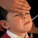
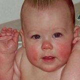
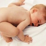
One of the characteristic symptoms of muscular torticollis is tilting the baby's head sideways or turning the chin in the opposite side of the head tilt. When trying to bring the head to a direct position nothing happens, but all because there is a strong strain of the sternocleidomastoid muscle. Upon examination, the doctor discovers that at the level of the middle third of the muscle, a spindle-shaped thickening is palpated and visualized, not welded to adjacent tissues and which is located in the abdominal muscle. With age, all symptoms appear stronger in the child, while the elasticity of the sternocleidomastoid muscle decreases. After the first year of life, asymmetry of the half of the facial skeleton and skull begins on the side of the head inclination.
At the age of three, the child is already showing a clear asymmetry of the face. Shoulders and shoulders are asymmetric, on the side of the torticollis they are slightly higher than on the opposite side. The neck on the side of the slope looks shorter. The sternocleidomastoid muscle is thickened compared to the same on the healthy side, with the exception of the middle third, where the dense spindle-shaped thickening is palpated.
Asymmetry of shoulder blades and shoulders is caused by the contraction of the trapezius and anterior staircase muscles. At an older age, children develop an upper thoracic and cervical scoliosis on the side of the torticollis.
Diagnosis
When a child is examined, the doctor clearly determines the asymmetry of the face on the side of the torticollis due to the narrower orbit and flattened superciliary arcs that are located slightly lower. In addition, the lower and upper jaws are flattened and underdeveloped. The ear from the side of the torticollis is located closer to the shoulder than on the healthy side.
It is very important that this disease is diagnosed even in the hospital or during the year after the birth of the child. This will increase the chances of a successful recovery. Also, timely detection of the anomaly will help prevent deformation of the skeleton of the face and head of the baby.
It is very important to distinguish congenital torticollis from other diseases: Klippel-Feil syndrome, additional cervical ribs, congenital accessory wedge-shaped cervical half-vertebrae, pterygium. In addition, the disease must be distinguished from vices and Grisel's disease, neck problems due to a generic craniocerebral trauma, spasmodic torticollis due to encephalitis and other forms of torticollis.
Treatment of torticollis
Treatment of torticollis will depend on the degree of the disease, as well as on the age of the child. Treatment can be conservative and surgical. Conservative treatment begins immediately after the umbilical ring is overgrown( at about two weeks of age the baby).With mild forms of the disease, doctors treat the patient with the help of such methods: massage, special styling, modes, Shantz collar and correction with a cotton-gauze pad.
The method of conservative treatment is simple. Both hands of one of the parents grasp the head of the baby, which lies on the back, then, without special efforts, you must gently tilt the head to the healthy side, while you must simultaneously turn the head to the sore side. Exercise should be completed on the area of the healthy half of the neck. After this, one of the parents should massage the muscles in the affected area. It is necessary to lightly press and palm the area at the level of compaction.
When the baby is laid to sleep, in the bed, you need to turn the healthy side of the neck to the wall. As a result, the baby will want to monitor everything that is happening around, and he will involuntarily stretch the affected sternocleidomastoid muscle.
In parallel with such treatment it is necessary to conduct courses of resorptive physiotherapy - electrophoresis( with potassium iodide) for 12-15 procedures, and massage courses. As a rule, after two or three such treatment courses, it is possible to achieve positive results. But doctors are obliged to warn parents about the possibility of a relapse, which can arise because the muscles on the affected side will lag behind in growth. Based on the foregoing, if a positive result is obtained, the parents are recommended to undergo four courses of physiotherapy with a massage in the first year of the baby's life, and then go through two or three courses of treatment in the second year of the baby's life.
Surgical treatment is prescribed for moderate to severe forms of the disease. Unfortunately, most often congenital torticollis is treated precisely by surgery, because conservative methods of treatment fail to achieve the desired result. As a rule, surgical treatment is carried out at 10-12 months of the baby's life. If you perform surgery after one year of life, this does not prevent the asymmetry of the facial skeleton. The operation is quite complicated, therefore, it is performed under anesthesia.
Depending on the degree of severity and the severity of the changes that occur in the sternocleidomastoid muscle, doctors prescribe one of the methods of surgical treatment: myotomy( muscle dissection) or plastic lengthening of the muscle.
Myotomy is performed under general anesthesia in the department of orthopedics. After the operation, the baby is made a special sticker and a cotton-gauze collar is applied. The next day after the surgery, the dressing is done and the neck is fixed with a plaster bandage in the hyper-correction position for a period of one month.
Plastic lengthening of the muscle is carried out for children aged 4-6 years and older. One of the advantages of such an operation is that it gives a more pronounced cosmetic result. After the operation, the patient is imposed for a couple of days correcting the cotton-gauze collar, then in the hypercorrection position - a plastic collar for two to three months.
Two weeks after the operation, the patient is prescribed a massage, physiotherapeutic procedures until full recovery. Three weeks after the operation, the patient is allowed to make light, active motions. If there is a relapse, the immobilization is continued for about another month. After removal of the immobilization, therapeutic exercises are directed to create new correct coordinated movements, as well as to restore the functionality of the elongated muscle. After surgery, the symmetry of the anterior triangle of the neck is restored.