Hamartoma
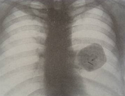 Hamartoma( Greek "hamartia" is a mistake) is the result of the pathological process in the tissues of a healthy organ. Hamartoma is a tumor-shaped nodal formation with a slightly pronounced tuberosity on the surface. They consist of the same tissues, where this benign tumor developed. Difference of tissues that make up the neoplasms, in the wrong location in the affected organ and in the degree of differentiation.
The lungs and the hypothalamus are most often affected, the hematoma of the liver, kidneys, and hamartoma of the breast are not ruled out. Dimensions of tumors can vary( 0.5-5 cm), they are usually not prone to rapid increase. Nevertheless, in the absence of treatment, the hamartoma is able to degenerate into a cancerous tumor.
Hamartoma causes
Hamartomas occur without any visible symptoms and are found randomly during X-ray examination for the presence of a very different disease, because the symptomatology of a hamart is similar to that of other diseases.
The risk group includes people 30-40 years old, while women are less susceptible to the emergence of this pathological process, in contrast to the male population( male lesions 2-4 times more).A newborn hamartoma can be detected with its inherent nature.
The main reasons for the development of hamartoma include:
- a violation of histogenesis and organogenesis in embryonic development( congenital hamartomas);
- a tumor as a consequence of the inflammatory process, during which occurs the appearance and proliferation of replacement tissue in an abnormal amount;
- in childhood, such neoplasms can occur due to injuries of the lungs, head or after various infectious diseases that contribute to the emergence of immunodeficiency;
- damage to the lung tissue;
- infection in the lower parts of the respiratory system;
- genetic predisposition;
- various gene mutations, the course of which is normally inadmissible;
is a harmful effect of tobacco smoke, chemical and radioactive substances falling into the soil, atmospheric air, water and polluting the environment. Such substances include formaldehyde, vinyl chloride, UV radiation, radioactive isotopes, etc.;
- the course of bronchopulmonary processes along with a decrease in immunity: the presence of bronchial asthma, chronic bronchitis, frequent pneumonia, tuberculosis, etc.
The presence of any of the above factors significantly increases the risk of a hamartoma.
Hamartoma symptoms
Distinguish between single and multiple forms of hamartomas. Also, depending on the prevailing type of tissue, the following tumors are distinguished in the hamartoma:
- chondro- matoma( in the tumor, the cartilaginous tissue is more localized);
- fibromatoma( the predominant tissue is fibrous, the tumor is most often found in the subcutaneous tissue);
- lipomatoma( in the composition of this form hamartom - fat tissue);
- angiomatoma( revealed in the vascular bed);
is a leiomyomatome.
In most cases, the presence of a hamart in the human body is asymptomatic. Detection of a hamartoma occurs randomly during an X-ray examination or fluorography of the organ where the given tumor is located.
X-ray picture of a hamartoma is similar to other diseases, therefore additional research should be carried out for an accurate diagnosis. On the roentgenogram, the hamartoma looks like a rounded shadow with clear contours, sometimes focal calcifications can be found against its background.
Over time, the size of the tumor usually remains unchanged, but with growth, it can cause the patient's discomfort in the area of the affected organ. Clarification of the diagnosis of a hamartoma is possible by means of a puncture biopsy if the neoplasm was found in the lung, or a bronchoscopy and a biopsy in the case of detection of endobronchial hamartomas.
With the development of a mild hamartomy, the symptomatology may include:
- hypoventilation of the affected lung;
- atelectasis of the corresponding lung and development of obstructive pneumonia;
- the appearance of hemoptysis in the event of destruction of the bronchus wall, after which the hamartomas become malignant;
- pain in the thoracic region, dry cough;
- wet cough with sputum separation, unpleasant sensations.
In general, the condition of the patient with this disease for a long time remains good.
The cerebral gamartoma is not uncommon, namely, the hypothalamus hamartoma. The flow of a hamartoma in this case is possible if:
- seizures( complex partial, generalized);
- epileptic fits of laughter;
- cognitive disorders: impaired attention, memory, assimilability of the material in the learning process, lack of assiduity, the presence of deviations in human behavior. It is more often found in childhood, in adults this symptomatology is not so pronounced;
- emotional disorders( depression, attention deficit disorder, autism, obsessive-compulsive syndrome).In children, in some cases, uncontrolled aggression is detected;
- violations in the functioning of the endocrine system. This is due to the fact that the hypothalamus takes part in the control of various organs;
- early puberty, which is possible without the presence of convulsions and other neurotic disorders.
In the case of a breast mammary gland, the course of the disease is also asymptomatic. The density of neoplasms depends on the ratio of fibrous, fatty, glandular tissues in them. Hence, it is possible to diagnose the hamartomy of the mammary gland upon palpation, during which a dense and mobile formation is detected. To study the tumor will help conduct mammography( on a mammogram it is well delineated with the presence of a thin, x-ray negative line around the entire formation or part of it).Ultrasound in this situation can be useless, since hamartomas are most often not recognized with this method of examination.
Hamartomas develop in the liver, with the lesion being more often exposed to its left share. For hematoma of the liver are characterized by:
- rapid or gradual increase in the abdomen for no apparent reason;
- abnormalities in the process of breathing, vomiting;
- a feeling of heaviness in the abdomen, nausea. This is due to the pressure of the hematoma of the liver on adjacent organs of the abdominal cavity;
- pain, indigestion;
- portal hypertension and ascites as a consequence of the pressure of the hamartoma on the portal vein.
Find the liver of the liver with its increase and already appearing symptoms. At the initial stages of development, hamartomas practically do not differ from healthy parenchyma of the liver. Echo- tomography and densitometry are useless here, but arteriography is able to detect a tumor due to its hypervascularization.
Hamartoma hypothalamus
Different brain hamartomas are quite dangerous for human health, since the brain is the head of all the processes of the body. Hypothalamus( department of the intermediate brain) is most susceptible to the appearance in it of a hamart. Although the size of this part of the brain is not large, it differs in structure in a complex way.
Under the control of the hypothalamus, many endocrine glands( pituitary gland, sex glands, thyroid gland, pancreas, adrenal glands, etc.) act, which in turn influence the constancy of the internal environment of the body. Disturbances in the functioning of the hypothalamus also lead to various endocrine abnormalities.
The hamartoma of the hypothalamus can develop as a benign tumor corresponding to the morphological classification of the ganglioncytome - a tumor consisting of elements of the nervous ganglia of the sympathetic system.
Diagnosis of the hypothalamus hamartoma, as well as the treatment of this disease, is complicated due to tumor localization and is based on the following survey methods:
- neurological examination( neurological functions are evaluated: sensitivity of the skin, vestibular apparatus, reflexes, functional state of the cerebellum);
- analysis for sex hormones;
- study of secondary sexual characteristics for premature maturation;
- application of visualization methods. Such methods include computed tomography( CT), as well as magnetic resonance imaging( MRI).The doctor receives, during the application of any of these methods, a layered image, with which it is possible to determine the size of the lesion, its location in the brain, the structure and shape. Experts believe that MRI is a more informative method of examining patients than CT due to the use of a magnetic field instead of X-rays.
Hamartoma of the lung
Hamartoma lung is a neoplasm with elements of the bronchial wall and parenchyma of the lung in its composition. The frequency of occurrence of hamart lung is from 0.25% to 0.4%.
Basically, a hamartoma consists of cartilaginous tissue( chondrogamarthoma), however, it is possible that other tissues of the lungs are present in the tumor( fatty-lipogamartoma, fibro-fibroid-fibrogamartoma).
The hamartoma of the lung is characterized by a rounded shape, even and sharp edges. Changes in the pulmonary tissue surrounding the neoplasm, and the root of the lung are absent.
As a rule, tumoral formations are located in the thickness of the parenchyma of the affected lung( intrapulmonary) near the visceral pleura or on its surface( subpleural).The small size of a hamart does not cause disturbances in the functioning of the lungs. A rare case is the location of a hamartoma within segmental, subsegmental bronchi. In this case, hamartomas can cause hypoventilation of the lung or its atelectasis.
In the diagnosis of hamartomas help:
- visit the therapist, pulmonologist, neonatologist;
- radiography of the lungs;
- appointment of a puncture biopsy;
- bronchoscopy.
The diagnosis is possible only after carrying out a fluorography or radiography of the lungs. Differential diagnostics with hamart with peripheral cancer, bronchogenic cyst, echinococcal cyst, limited pneumofibrosis, chronic abscess is also necessary. It should be borne in mind that prolonged observation of the patient is unsafe.
Indications for surgical intervention are available if the diagnosis is unclear for its clarification, radical removal of tumor-like formation. The operation itself consists in removing the tumor from the pulmonary tissue and further suturing. Less often, marginal resection is performed( removal of the tumor and a number of underlying tissues), segmentectomy, lobectomy.
Concerning the treatment of a lung gammarthoma, you should consult a doctor: an oncologist;Radiologist;A thoracic surgeon;Pulmonologist.
Hamartoma treatment
To date, several methods are used in the treatment of this disease, depending on the location of the tumor. When a brain, kidney, and lung brain is detected, surgical intervention is usually used. If there are contraindications to the operation, the patient is recommended to have regular follow-up.
With a hamartoma of a mild patient, regular examinations should be carried out, provided there is no increase in the tumor or other complications. If any, the patient is operated on removing the hamartoma. Surgical intervention can be:
- thoracotomy( tumor excision from lung tissue);
- fibrobronchoscopy( endobronchial hamartoma);
- vyluschivanie using videotorakoscopic techniques( the presence of a tumor in the subpleural region).
The treatment of the hypothalamus hamartoma includes:
- drug therapy( anticonvulsants).It serves as a symptomatic treatment, removing only the symptoms of the disease;
- surgical treatment( removal of a tumor in an operative way).In turn, the choice of the method of surgical intervention is made in accordance with the characteristics of the patient, the risk of postoperative complications;
- radiotherapy, and increasingly - radiosurgery( takes place at a high risk of surgical treatment, as well as when the tumor is hard to access).Radiation therapy includes gamma-knife technology, Novalis, a cyber knife. Radiosurgery, unlike radiotherapy, is a non-invasive technology, the essence of which is irradiation of a small dose of radiation with a hamartoma, along with the use of CT or MRI to monitor the direction of the radiation beam. Soft tissues around the tumor are exposed to a harmless dose of radiation, while tumor cells accumulate radiation, which is why the effect of radiation on them is greatly enhanced.