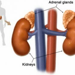
Kidney disease: nephroptosis

Causes of development of nephroptosis
In preserving the normal position of the kidney, its fat capsule is of great importance. The pericardial tissue, located around the kidney, holds the kidney's permanent position, limiting its movement. If the amount of fiber decreases sharply, the kidney begins to fall and can even turn around its axis. Sometimes the kidney turns around the vascular bundle, which makes up the renal artery and the renal vein.
Weight loss, especially severe, is one of the most common factors contributing to the development of nephroptosis. Also, this kidney disease also occurs with a decrease in the muscle tone of the abdominal wall. In this case, not only the kidney itself falls, but the rest of the abdominal cavity. Also, infectious diseases with the presence of connective tissue damage contribute to the development of nephroptosis. This tissue consists of ligaments and fascia, so their infection leads to a sharp drop in the organs. In women, nephroptosis occurs much more often than in men. The ratio of nephroptosis frequency in women is 1.7%, and in men - 0.1%.
Stages of development of nephroptosis
In the development of nephroptosis, physicians distinguish three stages:
- 1 stage. On it, the lowered kidney can be easily felt on the breath through the anterior abdominal wall, on exhalation the same kidney goes into the hypochondrium. In the normal position of the kidneys, they can be palpated only by very thin people, while the rest are not palpable.
- 2nd stage. When the patient is in the vertical position, the entire kidney leaves the hypochondrium freely, but when lying down, it returns to the hypochondrium. Sometimes it can be completely painlessly adjusted by hand.
- 3rd stage. The kidney leaves the hypochondrium completely in any position of the body, sometimes shifts into a small pelvis.
Already in the second stage, the kidney sometimes turns around its axis. The renal artery and vein thus bend, their lumen decreases, and vessels are also stretched. This significantly reduces the delivery to the kidney of arterial blood( ischemia) and causes difficulty in outflow from the kidney of venous blood( venous hypertension).At the third stage there is a steady bend of the ureter, and this can cause a violation of the outflow of urine. The disease nephroptosis, thus, in 2-3 stages leads to a significant pathology of blood supply to the kidneys, both venous and arterial. All this facilitates the development of kidney infections and the occurrence of pyelonephritis. The latter with nephroptosis often becomes chronic.
In the first stage, renal disease nephroptosis sometimes occurs imperceptibly. Occasional aching pain in the lower back can occur only occasionally. More often they begin to increase with physical activity and subside in a state of rest. Occasionally, there is a sudden transition from a prone position to a standing position.
In the kidneys, beginning with the second stage, the blood supply is disturbed, the urine stagnates, this can be accompanied by the appearance in the urine of red blood cells and protein.
In the third stage in the kidney region, the pain becomes permanent. The patient can have neurasthenia and depression due to these pains. Often, nephroptosis is accompanied by abnormalities in the work of the gastrointestinal tract, as well as a decrease in appetite. In the third stage, with severe ureteral bend during physical exertion, renal colic may occur. Often develops hypertension. This occurs especially often if the kidney artery is twisted and narrowed during nephroptosis.
Diagnosis of nephroptosis
Diagnosis of this disease is based on the patient's questioning and on the palpation of the moving kidney. Blood and urine tests are also being investigated. During the ultrasound examination, the mobile kidney is located in the patient's standing and lying position. The main method of diagnosing nephroptosis is the X-ray method. Excretory urography is also very important - a special study using contrast agents. It accurately shows the position of the lowered kidney. To clarify the status of kidney function, a radioisotope study is performed.
Treatment of nephroptosis
It is necessary to resort to operative treatment of nephroptosis if complications arise:
- intense pain that disrupts the life of the patient
- sharp decrease in functions of the lowered kidney
- chronic pyelonephritis
- arterial hypertension
- presence of blood in the urine
- hydronephrosis
Two weeks are administered to the operation of the patient. Anti-inflammatory treatment is performed in order to eliminate the possibility of spreading the infection during the operation.2-3 days before the surgery the patient begins to accustom to the lying position with the end of the bed, raised by 20 cm. In the same position, the patient is the first days after the operation. There are many ways of operative fixation of the kidney. The choice of the type of operation depends on the general condition of the patient and the surgeon's qualification. After the operation, the patient is prescribed anti-inflammatory treatment for two weeks, and if necessary, a mild laxative. After the operation, it is important for patients to limit physical activity for another 6 months.
General recommendations for nephroptosis
Implementation of these recommendations can significantly accelerate the treatment of nephroptosis:
- Before bed, put something under your feet, bringing them into a raised position;
- Do not lift anything heavier than seven kilograms with one hand. It's better to take them with you in a bag on wheels. At its absence, at least just distribute the weight on both hands equally;
- Complement exercise therapy with walking and swimming. Do an easy job at the household. Hard work at home should be excluded, as well as jumping and running.
You can not rely on physical exercises for nephroptosis in the following cases:
- If the pain is constant, and the kidney is lower than the fourth lumbar vertebra;
- If the pain does not decrease even when wearing a kidney bandage;
- If impaired kidney progresses;
- If due to the omission of the kidney, blood appears in the urine.
These signs indicate that surgery can not be avoided. The operation will be able to solve the problem completely: the kidney will be suspended on the muscle graft, it will always be in its place. Her normal mobility will be preserved, her normal work will be restored.


