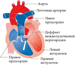
Tetrada Fallot
 The tetralogy of Fallot is a severe congenital heart pathology, which is characterized by underdevelopment of the terminal section of the right ventricle with a displacement of the septum to one side. As a result of such changes, four major defects are formed: hypertrophic changes in the myocardium, aortic destruction, subaortal defects of the interventricular septum, and stenosis of the outlet of the right ventricle.
Clinically, such a pathology as the tetralogy of Fallot is manifested by a delay in physical and mental development, early cyanosis of the skin( sometimes cyanosis occurs already from the first months after birth, but more often by a year or even later), shortness of breath or severe dyspnea-cyanotic attacks, frequent fainting, Nausea, dizziness and constant weakness.
The tetralogy of Fallot causes
The development of the tetralogy of Fallot can be caused by a violation of the full development of the heart for 2-8 weeks of fetal embryonic development. In addition, infectious diseases transferred by a pregnant woman, such as measles, rubella or scarlet fever, also lead to the development of cardiovascular malformations. Taking drugs, drugs, alcohol, smoking and exposure to various harmful environmental factors can also lead to the development of pathology. Equally important is the influence of heredity on the development of the fetus.
The development of the tetralogy of Fallot in children with the syndrome of Amsterdam dwarfism( Cornelia de Lange), which includes such pathological conditions as: "face of the clown";Gothic sky;Deformation of the spine and thorax;Atresia of the hoan;strabismus;Atrophy of the optic nerves;Deformation of the auricles;Hypertrichosis;myopia;Absence of some fingers;Severe defects of internal organs;Low birth weight;Small feet and palms.
Due to the fact that the aorta departs from both the left and right ventricles, with severe stenosis or significant defects during systole, almost all of the blood from the ventricles enters the aorta and in very small volumes into the pulmonary artery. As a result, right ventricular failure develops. In the presence of moderate stenosis, the acyanotic form of the tetralogy of Fallot is manifested. With an increase in the severity of stenosis, there is a cross-discharge of blood, and then from right to left with a transition to the pronounced stage of the acyanotic form.
The Fallot form of the
form. To date, there are four main forms of obstruction of the right ventricular outflow duct: embryological, hypertrophic, tubular, multicomponent.
Embryological form. This form of the disease is characterized by the displacement of the cone septum anteriorly and to the left. The zone of maximum development of stenosis corresponds to the muscular ring. In this case, the fibrinous ring does not deform.
Hypertrophic form. Obstruction in this case is characterized by the same displacement of the septum as in the embryological form, but in addition, its pronounced hypertrophic changes in the proximal segment are noted. In this case, the zone of maximum development of stenosis is at the level of the output section of the right ventricle and the delimiting muscle layer.
The tubular form. Obstruction in the third type of tetralogy of Fallot is caused by the unevenly formed division of the common arterial trunk. As a result, the pulmonary cone becomes shortened, narrowed and hypoplastic. Also this form is accompanied by hypoplasia of the fibrinous ring and stenosis of the pulmonary artery.
Multicomponent form. The cause of the development of obstruction in this form of the tetralogy of Fallot is the elongation of the conical septum.
The degree of severity of hemodynamic disorders manifests itself in the form of obstruction of the ventricular duct of the right ventricle. With severe stenosis, the flow of blood from both ventricles is noted mainly in the aorta and in a very small volume in the pulmonary artery, and this condition is accompanied by arterial hypoxia.
With moderate stenosis, peripheral resistance is greater in the output tract, and therefore a left-right ventricular outflow from the ventricles develops, which subsequently leads to the acyanotic form of the disease. In another way, this condition is called the pale form of the tetralogy of Fallot. However, over time, the "pale" form grows into a "blue" form, this is due to the development of cross and venoarterial discharge of blood.
Tetrada Fallot symptoms
Depending on the time of development of the initial symptomatology, five main periods of the manifestation of the tetralogy of Fallot are distinguished: it is an early cyanotic stage( the appearance of cyanosis in the first months of life or closer to one year of life), the classical stage( the appearance of cyanosis at the time of the child's movement), the severe stage(Attacks of dyspnea with obvious manifestations of cyanosis), late cyanotic stage( the onset of symptoms is not earlier than 6-10 years), acyanotic( pale) stage of the disease.
Most often, with a disease such as the tetralogy of Fallot, the following complaints and manifestations occur: developmental delay;dyspnea;Severe attacks of dyspnea with manifestations of cyanosis;Central cyanosis;loss of consciousness;Convulsions.
Objectively during the survey, it is noted:
- pallor of the skin or cyanotic manifestations;
- a symptom of "drum sticks" or "watch glasses";
- strengthening of the first tone of the heart;
- the position of the patient squatting after small loads;
- attenuation of the second tone above the pulmonary artery;
- the presence of systolic or systolic diastolic.
Tetrada Fallo Diagnosis
To confirm the diagnosis of "tetralogy of Fallot" it is necessary to collect a complete history of the disease, a clinical picture of the manifesting symptoms, conduct an ECG and chest X-ray.
Instrumental diagnostic methods
ECG( electrocardiogram): during this study, signs of hypertrophy or overload of certain parts of the heart, AV blockade and syndrome of weakness of the sinus node are noted.
Chest x-ray: for Fallot tetrad, depletion of the pulmonary pattern, swelling of the right or left ventricle arches, with a pronounced combination of open arterial defect is characteristic. In young children, the heart on the roentgenogram has the form of a "shoe".
Echocardiogram: this method of diagnosing the tetralogy of Fallot allows to estimate the sizes of the cavity of the right ventricle, the degree of narrowing of the outlet duct, to determine the form of the pathological process( embryological, hypertrophic, multicomponent or tubular).The same method is performed for the purpose of diagnosing the aortic valve for the presence of stenosis, since in time the undetected problems associated with the development of the valve after treatment of the defect can lead to rapid decompensation or pulmonary edema. And also using this method, the diameter of the fibrous rings is determined and the general state of their wings is examined.
Cardiac catheterization: cardiac puncture is performed when it is necessary to determine the oxygenation of blood in the aorta, measure the pressure in the right ventricle, or the pressure gradient between the right ventricle and the pulmonary artery.
Coronary angiography: this method is used to determine the anatomical type of heart disease, to diagnose concomitant developmental anomalies and to determine the localization of coronary arteries and major vessels.
Tetrada Fallot treatment
To date, there are two main methods aimed at treating tetralogy of Fallot: surgical treatment and urgent therapy( treatment of dyspnea-cyanotic attack).
Urgent therapy for
Newborns with severe manifestations of cyanosis due to the closure of the arterial duct are immediately injected with infusions of prostaglandin E1 at 0.05-0.10 μg intravenously. This manipulation is carried out in order to open the arterial duct in the shortest possible time.
During severe hypoxic attacks, the child is given a position where the legs are as much as possible pressed to the chest, the older children just squat and the attack passes, and morphine is prescribed 0.1-0.2 mg / kg IM.
The dyspnea-cyanotic attack is stopped with oxygen therapy, 1% tri-pleidine solution intramuscularly at 0.05 ml in the first year of life or Nicomedin at 0.1 ml. If there is no effect, intravenous crystalloids should be administered. When acidosis is introduced 4% solution of sodium bicarbonate, 5% glucose solution with the addition of insulin, rheopolyglucin and aminophylline. If there is no effect, an immediate superimposition of the aorto-pulmonary anastomosis is performed. It is also possible to use sedative and / or vasodilator drugs.
To a specific method, the treatment of tetralogy of Fallot is a slow intravenous injection of beta-adrenoblockers at 0.1 mg / kg, and then applying them as tablets at 1 mg / kg per day.
Patients with a disease such as the tetralogy of Fallot are categorically contraindicated in the appointment of Digoxin, since it increases ionotropic functions of the myocardium and increases the risk of developing a spasm of the infodibular portion of the right ventricle.
Surgical treatment
Contraindication to the treatment of tetralogy of Fallot by surgery is an irreversible pulmonary hypertension.
First of all, the choice of an operative method of treatment depends on the anatomical structure of the heart defect. It can be both palliative and radical. The palliative method of treatment of the tetralogy of Fallot involves the imposition of aorto-pulmonary anastomosis, and the radical one - the elimination of stenosis with subsequent plasty of the interventricular septum.
Palliative surgery is prescribed if the shortness of cyanotic attacks can not be stopped with medication for a long time, as well as with anatomical features of the human body.
The most commonly used method is fixation of subclavian-pulmonary anastomosis. The essence of the operation is that the subclavian artery is connected to the pulmonary artery by means of special synthetic implants. Experts recommend immediately to carry out radical methods of treatment.
Radical surgery is mainly performed at the age of 6 months to 3 years, as at a later age the chances of a full recovery are reduced. This method of treatment involves carrying out the plastic of the interventricular septum and eliminating the obstruction of the excretory part of the right ventricle. A complete correction consists of closing the defect with a patch and expanding the duct.
All patients diagnosed with the tetralogy of Fallot, regardless of whether a correction was made, must take preventive measures before going to the dentist, in order to avoid the development of bacteremia.
Tetrada Fallo forecast
Specific postoperative complications: hypofunction;Thrombosis of anastomoses;Ventricular arrhythmia;AV blockade;Aneurysm of the right ventricle;Infective endocarditis;Pulmonary hypertension. Dyspnea-cyanotic seizures often pass independently after 3-4 years.
Postoperative lethality today is less than 4%, with uncomplicated form of the disease. If you do not operate such a pathology, then up to 5 years of age survive slightly more than 55%, and up to 10 years - 25-30%, in very rare cases patients with a tetralogy of Fallot live up to 75 years.