Aortic stenosis
 Aortic stenosis is a disease related to heart disease. By the word vice means organic damage to the structure of the heart valve or the valve opening. The causes of the appearance of such diseases are different, from congenital pathologies and to the consequences of infective endocarditis.
The peculiarity of the group of diseases, which includes aortic stenosis, is the appearance of serious hemodynamic disorders. Since normal blood flow is disturbed, the heart has to pump large volumes of blood and cope with higher intracardiac pressure than if it coped with those in norm. As a result, a number of injuries occur in the work of individual organs.
You also need to know that aortic stenosis has one important feature - it has an extremely slow manifestation. This is due to a stronger left ventricular muscle. He copes longer with the effect of increased pressure, which occurs in it with aortic stenosis.
Stenosis of the aortic valve
Stenosis in medicine is understood as constriction. Stenosis of the aortic valve is characterized by a persistent constriction of the aortic aorta. It can be either acquired( under the influence of certain diseases) or congenital( a genetically caused defect in the valves of the aortic orifice and consequence of this is congenital aortic stenosis).
For localization of damage, stenosis of the aortic valve is subdivided into subvalvular and valve stenosis. The most common type of aortic stenosis is, of course, acquired valvular aortic stenosis. When the medical literature mentions the diagnosis of "aortic stenosis", by default, it is specifically congenital valvular aortic stenosis.
Aortic stenosis causes
One of the most common causes of aortic stenosis is rheumatic fever. Under the influence of the causative agent of rheumatism B hemolytic streptococcus, the commissure melts( connective tissue valve supports), then the valves of the aortic valve splice and fibrotic. After this, calcifications are deposited on the valves and the size of the aortic aperture is considerably narrowed. As a result, aortic stenosis develops.
Also, the appearance of aortic stenosis is caused by age-related changes: the appearance of calcium salts on the aortic valve, subsequent fibrosis and mobility impairment.
It is also important to understand which pathological processes trigger aortic stenosis in the heart. These processes are extremely important for proper perception of the clinical picture and the purpose of the current treatment regimen.
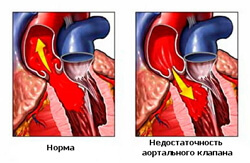
Normally, the size of the aortic aperture in an adult is about 4 cm. When aortic stenosis appears, this hole narrows. As a result, the aortic aperture has a much smaller area than in the norm. Reduction of the aortic aperture area leads to complication of the passage of blood flow into the aorta from the left ventricle. In order to change and remove this difficulty and not to disrupt the process of normal blood supply of the body, the heart tries to increase the level of pressure in the left ventricular chamber. The pressure increases at the time of systole of the left ventricle - the moment when the blood is throttled to the aorta by heart. In addition, the heart automatically lengthens the systole time. Thus, the heart tries to "force" to squeeze blood through the narrowed aortic mouth, affected by aortic stenosis, and increase the time of blood transfer to the aorta. The heart triggers these mechanisms for the sole purpose of ensuring a normal discharge of blood into the aortic system.
The effect of increased pressure in the left ventricular chamber does not go unpunished for the heart. In response to an increase in systolic pressure, hypertrophy of the muscular layer( myocardium) occurs in the left ventricle. This develops so that the myocardium can cope with a high pressure gradient and ensure that it releases a volume of blood that can supply blood to the organs that need it. But the increased muscle mass of the left ventricle is much worse relaxed and stretched. Because of this during diastole( relaxation of the left ventricle) diastolic pressure rapidly increases.
Normally, the heart performs the following cycle:
1. Atrial systole: due to muscle contraction, the blood is pushed into the ventricles. Then the atria relax, and their diastole sets in.
2. Ventricular systole. During contraction of the ventricular myocardium, the blood flow goes to the pulmonary veins from the right ventricle, and into the aortic system from the left. And then he rushes into the circulation.
3. A common diastole.
It is important to understand that when one phase of the heart has one phase of the cardiac cycle, then in the other department it passes completely the opposite. So, when in the atria of the systole, then in the ventricles at that moment there will be a diastole.
Thus, the high pressure in the left ventricle chamber at the time of its relaxation will disrupt the process in which the left atrium will push the flow of blood into the left ventricular chamber. In simple words, the left atrium will not be emptied completely, and there will be a certain amount of blood in it. Therefore, the left atrium increases the number of its contractions, so that there is no "excess blood" left in it.
But, despite the fact that the myocardium of the left ventricle is strong enough, even it can not always cope with the increasing pressure. At a certain point, the ventricle ceases to resist pressure and is dilated( stretched) under its action. In the stretched ventricle, the pressure continues to grow and already affects the left atrium. Unable to fight with high diastolic pressure in the chamber of the ventricle, being full of blood, the atria also stretch. High left atrial pressure affects the pulmonary veins and a phenomenon such as pulmonary hypertension appears.
As a result, the appearance of aortic stenosis leads to the following pathological conditions:
1. The stretched left ventricle can no longer discharge normal blood volume, which means that the left ventricular function may fail.
2. As a result of the prolonged existence of aortic stenosis, which is no longer accompanied by any compensatory processes, the right heart can be affected. As a result of the effect of increasing pressure, mitral-aortic stenosis will develop first in the left ventricle, then in the left atrium, its subsequent exposure to the pulmonary veins. It will be characterized by the presence of aortic stenosis and insufficiency of the mitral valve, which will appear due to the dilatation of the right heart.
It is believed that the time for which the myocardium of the left ventricle stops coping with high systolic pressure and finally spreads, causing subsequent changes characteristic of aortic stenosis, averages 4 years.
By the way, in the clinic of aortic stenosis there is a so-called concept of "critical stenosis".This reduction of the aortic valve aperture to 0.75 cm 2. With this variant of aortic stenosis, the patient will quickly develop pulmonary edema and heart failure.
And if the aortic valve aperture has an area from 1.2 cm to 0.75 cm, this stenosis will be called moderate aortic stenosis.
Accordingly, with narrowing of the aortic valve opening area in the range from 2.0 to 1.2 cm, this stenosis will be called insignificant.
Aortic stenosis symptoms
As described above, patients usually do not complain of aortic stenosis for quite some time. For a long time he is in the stage of compensation. Complaints of patients, on any unpleasant sensations appear, when the aortic valve aperture is already destined almost half.
The first "bells" for the possible presence of aortic stenosis are syncopal or syncope. Their manifestation is explained in the following mechanism, which accompanies aortic stenosis. The fact is that with aortic stenosis, a mechanism of "fixed ejection" appears. It consists in the fact that during exercise the heart can not increase cardiac output due to the narrowing of the aortic valve aperture. Due to the lack of adequate blood supply, with physical exertion, dizziness, nausea, and weakness appear in patients with aortic stenosis. At late degrees of stenosis, temporary loss of consciousness may even appear.
Also, signs of aortic stenosis may manifest in cardiac ischemic pain. This clinical manifestation is explained by the fact that in the valves of the aorta valves coronary arteries begin, which enter the blood supply system of the heart. Because of a violation of the level of normal pressure between the left ventricle and the aortic aperture, there is a decrease in the flow of blood into these arteries. As a consequence, the blood supply to the heart is not enough, which means that myocardial ischemia and its clinical manifestations, heart pain, will appear.
The third subjective complaint for aortic stenosis will be shortness of breath. Arising as a consequence of a disturbance in the functioning of the left ventricle, first in diastole, and then in systole, dyspnea can turn into left ventricular failure. It will show up with a cough with abundant, foamy, pinkish phlegm. It is foamy sputum with a pinkish color - this is a bright sign of stagnation in the pulmonary circulation. Pink color in sputum is due to a slight transition of red blood cells from the pulmonary artery system to the alveoli, which are tightly surrounded by pulmonary vessels.
But finally to confirm the diagnosis of aortic stenosis is possible only with a full examination of the patient. This is the only way to identify a number of symptoms that are characteristic of stenosis of the aortic aorta.
First of all, pay attention to the appearance. With aortic stenosis, the pallor of the skin will be noted. This is due to all the same impaired and reduced outflow of the left ventricle.
Since the left ventricle with aortic stenosis has an even higher muscle mass and shrinks faster, it is not difficult to determine the strong apical impulse and the characteristic concussion of the entire atrial region. Most often this concussion is in resonance with cardiac contractions.
If you put your hands in the second intercostal space on the right circumcostal line( this is the point where the aorta is usually dried), you can feel the trembling of the fingers that will appear at the time of contraction of the left ventricle. This tremor is the result of the flow of blood through the narrow aortic estuary. And the more narrow the aorta, the brighter and better this jitter or "purring" will be determined.
If you start a pulse test, then a patient with aortic stenosis will have a rare, small filling. The more rare acronyms are explained by the second compensatory mechanism, which will start the heart - by increasing the frequency of contractions of the left ventricle.
And if you examine the blood pressure, then the presence of aortic stenosis will be reduced in comparison with normal systolic( "upper") pressure.
But, undoubtedly, the most correct clinical sign of aortic stenosis will be a certain picture, which the doctor will listen with a stethophonendoscope.
When aortic valve is dried, a systolic, coarse noise can be easily detected by a stenotic aortic stenosis. The reason for its occurrence is the same as in the "jitter", which has already been described above. Blood goes through the narrowed part of the aorta, its "twists" arise and noise is heard. Like jitter, this phenomenon occurs at the time of contraction of the left ventricle( that is, its systole).
In addition, there are other interesting changes in the operation of the valvular apparatus of the heart, which can be listened to. Since a small amount of blood is sent to the large circle of blood circulation, the second tone( and it is the tone of the left ventricle diastole, and is obtained when the three valves of the aortic valve is slammed) is much weaker than normal. A small amount of blood slams the aortic valve with little force. Sometimes this second tone can even be difficult to listen to. The reason for this is very simple: with aortic stenosis, the valves are most often fibrous, which means they are inactive and, again, are slammed with much less strength and sound.
While the diastolic pressure did not begin to increase in the left ventricular chamber, the 1st tone( tone of purity) will sound relatively normal. But since the duration of the left ventricle systole is increased, the doctor with a musical ear will be able to listen to a slight decrease in the sonority of the systolic tone.
In addition, when there is decompensation, and pressure during diastole increases - in the region of the cardiac apex appears 3rd and 4th tones. These tones do not manifest themselves during the normal functioning of the heart valve apparatus, and therefore, are considered pathological. These tones will tell the doctor that the left atrium has already been involved, and it is struggling to push the blood into the left ventricle, where high diastolic pressure interferes with it.
When a pathological process is started, the clinical symptoms go beyond the heart and are located in those organs whose blood supply is subject to disturbance, with that or other prophet. With aortic stenosis, the lungs most often suffer. If you listen to them with a stethophonendoscope, you can hear wet bubbling rales - this is a sign of the presence of fluid in the lungs. Also, tapping the lungs, if there is liquid in them, you can hear how the percussion sound becomes shorter and less sonorous.
Aortic stenosis treatment
Treatment of aortic stenosis includes surgical and conservative methods. In conservative methods, attention is focused on eliminating the consequences of blood flow disorders, cardiac arrhythmias and the prevention of the occurrence of infective endocarditis.
The first to try to eliminate the phenomenon of stagnation in the pulmonary circulation. Assign diuretics( most often use Furosemide).It is important to prescribe them, taking into account all clinical, instrumental and subjective data and apply with great care.
At cardiac arrhythmia, cardiac glycosides are prescribed( Digoxin).
Potassium preparations are also quite widespread in the treatment of aortic stenosis.
To improve relaxation of hypertrophied myocardium, B-blockers or antagonists of calcium blockers( especially those belonging to the Verapamil series) are used.
The use of a group of nitrates in aortic stenosis is contraindicated. Nitrates reduce cardiac output and minute blood volume. This can easily lead to a drop in blood pressure to a critical level.
Most often, conservative methods of treatment are combined with surgical ones: they are used for preoperative preparation of the patient and in the postoperative period.
But the main method of treatment for aortic stenosis are surgical methods of treatment. They depend on the degree of decompensation of the defect, various disorders that are the consequences of the defect and the contraindications present.
The most common use of aortic valve replacement or balloon valve plastic.
The main indications for surgical correction of aortic stenosis are:
1. The presence of satisfactory function of the myocardium.
2. If the systolic pressure gradient in the aortic valve region is higher than 60 mmHg, Art.
3. On the cardiogram, there are signs of increased left ventricular hypertrophy.
In turn, the main contraindications to the operation will be:
1. Pressure gradient on the aortic valve above 150 mm from st.
2. Severe dystrophic changes in the left ventricular myocardium.
Aortic stenosis operation
One of the most frequently used surgical procedures is the artificial prosthesis of the aortic valve. If, however, minor changes are revealed in the valve flaps with aortic stenosis, then it is more expedient to confine oneself to less voluminous surgical intervention: operative separation of the spliced aortic valve flaps.
The operation for the artificial replacement of the tricuspid aortic valve is carried out by first connecting the patient to the cardiopulmonary bypass apparatus. That is, the heart is completely cut off( "de-energized") from the total blood flow.
After starting the artificial circulation, the aorta dissects and the aortic valve is inspected, then it is removed. The size of the aortic valve aperture is measured, its dimensions are checked against the implant, which is located immediately in the operating room. Having convinced that the implant corresponds to the size of the hole, it is sewn. Then the surgeon checks the operation area for integrity. After this, the functional capacity of the new prosthetic valve is checked. Then possible air bubbles are removed, since their presence with natural blood flow to the heart can result in embolism and death. After all this, the chest is closed and sewn up.
After surgery, much attention is paid to the prevention of postoperative complications. Most afraid of the emergence of postoperative infective endocarditis. That is why patients after an operation to correct aortic stenosis are on therapy with broad-spectrum antibiotics. The second complication, which is most feared is thromboembolism. Because of this, patients after such surgery for a long time take anticoagulants and antiaggregants. Most often they like to use Aspirin and Heparin.
During the prosthetic operation, various types of implants are used: ball or disc prostheses made of artificial materials or biological implants, taken from the biological material of the patient himself. Of course, best of all, it is the second subset of prostheses. Since they are taken from the patient's body, this reduces the risk of rejection of a new valve in patients with aortic stenosis.
It is important to understand that the earlier the new valve is implanted, the better results can be obtained. If you perform an operation in the early stages of aortic stenosis, the risk of postoperative complications will be much lower and postoperative rehabilitation for the patient will be easier.