In what situations are MRI or CT of the brain assigned?
Contents:
- What is CT?
- What is an MRI?
- Features of the procedure
- Conclusion
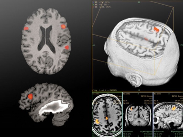 Patients are interested in what is better for examining the brain: CT or MRI?Both studies are non-invasive and show well the pathologies of the central nervous system. What is the difference between MRI and CT of the brain? At what pathologies it is better to appoint CT, and at what - MRT?Each of the methods has its advantages and disadvantages, therefore both methods are popular for examining the central system.
What is CT?
Computerized tomography of the head is a method of investigation in which X-rays are used. A computer tomograph consists of a ring in which a couch with a patient is placed, scanners and a computer receiving signals. In the ring, emitters are built in, emitting X-rays at a certain angle in order to take a snapshot.
The structures of the brain reflect or absorb this radiation at different intensities, which is fixed by scanners built into the tomograph tube. From the scanners is information to the computer in the form of black and white pictures in a section. There is a possibility of 3D-modeling.
CT is more informative than ultrasound. The examination is prescribed with:
- permanent and severe pain;
- dizziness, loss of balance, regular fainting, tinnitus;
- craniocerebral injury;
- suspected of having a stroke;
- problems with teeth, intraosseous sinuses( frontal sinus, sinusitis, sphenoiditis, etmoiditis);
- suspected of the presence of neoplasms of the central nervous system.
 Find out what duplex scanning of the vessels of the head and neck is and how the study is done.
All about the NSH of the brain of newborns: the procedure for conducting, analyzing the results of the NSH.
The difference between CT and MRI of the brain is that X-rays in computed tomography make it possible to see better bone formation than soft tissue and vessels. If the procedure is carried out without contrasting, it lasts a short time - a few seconds. This is its advantage over magnetic resonance imaging of the brain.
Especially this plus is important when examining children and people with claustrophobia, who are afraid of enclosed spaces. CT scan better sees calcified formations, as well as long-standing post-stroke changes.
When contrasting, the duration of the examination can reach 30-50 minutes, which is comparable in duration with magnetic resonance imaging. This may require sedation or anesthesia in children or those with claustrophobia.
The cost of the survey is slightly less than that of the magnetic resonance method, the difference is usually one thousand rubles. Conducting CT in Russian medical institutions costs about 3000-4000 rubles.
Another advantage - for CT, the presence of metal-containing tattoos and other metal objects( skull plates) is not an obstacle. Minus - X-ray radiation affects the body not in the best way, therefore, for pregnant women CT is contraindicated throughout the period of gestation.
What is an MRI?
Magnetic resonance imaging is often prescribed after ultrasound. This method uses a magnetic field of a certain frequency. Magnetic resonance tomograph is similar to computer tomography. It also has scanners for receiving a response signal from the tissues.
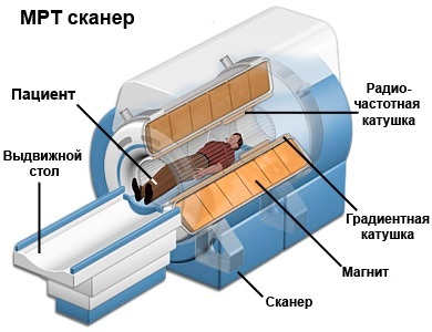 The ring in which the patient is immersed on the couch is represented by a radio-frequency coil from which waves emanate. Inside the ring, a powerful permanent high-voltage magnetic field is created - up to 3 Tesla. Under such conditions, active intermingling of hydrogen atoms begins in the human tissues, which is fixed by scanners of the magnetic resonance tomograph. The information from the detectors in the ring comes to the computer in the form of snapshots and 3D images.
Features of the
procedureThe duration of the procedure is about 20-30 minutes, with a contrast of about 50 minutes. This presents a particular problem when examining children and people with claustrophobia. It's hard for a child to lie for a long time without moving, and with claustrophobia there is a fear of closed spaces, which can lead to unpleasant consequences. Therefore, in these groups of individuals, MRI is not used, or is performed with sedation or under anesthesia.
Restriction in the study: the presence of metal objects in the body( skull plates), pregnancy up to 12 weeks. Contrasting is contraindicated in cases of hematopoietic anemia. Contrast substance introduced inside the vessels of the head, has some toxic effect on the body, as in CT.
Important! Advantage of magnetic resonance tomography before CT is the absence of X-ray radiation. This allows you to conduct a survey as often as required.
Magnetic resonance imaging is indicated with the following symptoms:
- Vertigo and headache, which are of a regular nature or high intensity.
- Noise in the ears.
- Suspicion of neoplasm or cerebral hemorrhage.
- Hearing loss, vision, paralysis.
- Symptoms of multiple sclerosis.
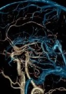 Methods for diagnosing the vessels of the head and neck: how to simultaneously examine the vascular system of the head and neck.
Did you know that angiography of cerebral vessels is an effective diagnostic method.
Find out what is dopplerography of the vessels of the head and neck: indications for carrying out.
Magnetic resonance imaging is prescribed more often, as it is better than CT to "see" soft tissue. However, long-term changes after a stroke on MR images are worse than in CT images.
The magnetic field has some negative effect on the body and red blood cells, since they contain iron. However, these changes are insignificant. The price of this method in Russian clinics is about 4000-5000 thousand rubles.
Conclusion
CT and MRI of the brain are the most reliable methods of diagnosing and visualizing diseases of the central nervous system. They are appointed, as a rule, after ultrasound examination to clarify the situation, clarify the diagnosis. The doctor who prescribes this or that research takes into account the nature of the pathology: what tissues are affected - soft or bony formations.
MRI or CT of the brain? If there is a suspicion of inflammatory processes in the paranasal sinuses, temporal bone( mastoiditis), eyeballs, it is better to do computed tomography. However, soft tissue and vascular pathologies are better detected on MRI.
write the question in the form below:


