Preparation and conduct of MRI of the prostate( with and without contrast)
MRI, or magnetic resonance imaging, , is a non-invasive technique that provides reliable information about the condition of the organ. A layered image of the prostate can reveal abnormalities of development, abscesses and other pathological changes. Of great importance is MRI in the diagnosis of cancer and other neoplasms of the prostate.

Magnetic resonance imaging - the most informative study of prostate
Content
- 1 Indications
- 2 Contraindications
- 3 Preparation for the procedure
- 4 Technique
- 4.1 MRI spectroscopy
- 4.2 MRI with contrast
- 4.3 multiparametric MRI
- 5 Benefits
Indications
Magnetic Resonance ImagingIs far from being a routine procedure. Manipulation is performed to clarify the diagnosis after a previous laboratory and instrumental examination. MRI is usually preceded by examination of the urologist, blood and urine tests, ultrasound.
MRI of the prostate gland is performed with states :
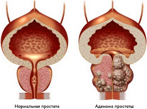
- suspected of prostate cancer or adenoma;
- prostatic abscess;
- prostatitis;
- organ development abnormalities;
- definition of the zone of the forthcoming surgical intervention.
In most cases, the procedure is performed with a suspected cancer or adenoma of the prostate. Sighting allows to detect even small foci of carcinoma, to reveal metastases to regional lymph nodes and to clarify the stage of the disease. MRI also monitors the effectiveness of treatment after resection of the prostate or complete organ removal.
Contraindications
MRI is not carried out if the patient has non-removable metal implant or implanted electronic devices, including:
- implants joints;
- implants of the inner ear;
- stents of blood vessels;
- artificial heart valves;
- pacemakers;
- metal pins( including those used for the installation of dental crowns);
- metal elements in the body( fragments of bullets, etc.).
For relative contraindications applies:
- claustrophobia( fear of enclosed spaces);
- patient weight more than 100 kg.
In these situations, the procedure is performed on open type devices.
Preparing for
procedure Before performing the test, the physician explains in detail the technique of performing it. Particular attention is paid to the behavior of the patient during the procedure and the probable sensations during MRI.To obtain a reliable result recommended :
- Within 3 days prior to MRI abandon the use of products that provoke the formation of gas in the intestine.
- Restrict the use of heavy meals 12 hours before the procedure.
- You can not eat any food 4 hours before the test.
- Carry out a cleansing enema in the morning before the MRI( when using a transrectal sensor).
- remove from the body and lay out of the pockets of all metal objects. Jewelery, watches, pins, pens, etc. metallic piercing and dentures also need to be removed.
During the procedure, the patient can be in his usual clothes, but only if it does not contain metal parts( zippers, buttons).In many clinics, the patient is offered to change into a sterile set of clothes.
Technique for running
There are two types of MRI devices: closed and open type. The classical apparatus is a large cylindrical tube surrounded by a magnet. The patient is on a movable table, which is placed inside the magnetic field. In open-type devices, the magnet is located only on the sides of the patient. This method is recommended for people suffering from claustrophobia( fear of closed spaces).
The essence of MRI is to obtain a layered image of the organ. All information is displayed. Such a technique allows to identify tumors and other prostate formations. With the help of MRI it is possible to detect even small enough foci, as well as to assess the blood flow in the suspected zone.
During the MRI, the patient should remain completely immobile for a while. If this can not be achieved, the doctor may suggest another method for examining the prostate gland.
There are several options for conducting MRI:
MRI spectroscopy
The procedure is performed using an endorectal coil. The coil is placed in a disposable cuff, lubricated with a lubricant and carefully inserted into the rectum. In the intestine, the cuff is inflated, which allows it to be held in a fixed position during the examination. At the end of the procedure, the reel is removed. All manipulations take about 20-30 minutes.
MRI with contrast
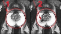
MRI with contrast:
1 - healthy prostate;
2 - prostate cancer
The procedure allows you to identify atypical cells. It is known that the cancer tumor contains much less water than in normal tissue. Contrast substance, entering the prostate tissue, transmits this visual difference to the screen. MRI using contrast is prescribed for suspected cancer, as well as for monitoring the effectiveness of the treatment.
The scheme of MRI with contrast is not too different from the usual procedure. Before the beginning of the study, a contrast agent is injected into the vein, after which a series of images is taken. If there is an undesirable reaction to the drug( skin rash, itching), it is necessary to inform the doctor about it. The duration of the procedure is from 45 minutes to an hour.
Multiparameter MRI
To date, multivariate MRI is considered the most effective method of detecting prostate cancer in the early stages. The procedure allows you to assess the nature of blood flow in tissues, determine the concentration of chemicals and the rate of movement of water. With the help of a progressive technique, a doctor can detect small foci of a cancerous tumor and accurately identify the zone of necessary surgical intervention.
MRI of the prostate is often combined with a biopsy. Under visual control, the doctor accurately takes the desired area of prostate tissue. This approach significantly improves the accuracy of diagnosis and increases the chances of detecting cancer at an early stage.
Advantages of
The MRI technique has unique advantages of over other methods:
- High accuracy of research and good visualization: the opportunity to see a cancerous tumor at early stages of its development.
- Painlessness: the patient practically does not experience unpleasant sensations during the procedure.
- Safety: no contact with skin is required, ionizing radiation is not used.
- Fast result: a diagnosis can be made immediately after the procedure.
Magnetic resonance imaging is one of the leading methods for diagnosing cancer and other dangerous prostate diseases. The technique allows not only to detect the tumor, but also to assess the extent of its spread. Timely diagnosis of cancer significantly facilitates treatment and increases the chances of a favorable outcome of the disease.
Recommended for viewing: