Duplex scanning of vessels of the head and neck
Contents:
- What is duplex scanning?
- How is the duplex study of the vessels of the head and neck
 Duplex scanning of the vessels of the head and neck is an ultrasonic study of vessels that are extracranial( vessels of the neck) and intracranial( head vessels).This is a modern diagnostic method that allows you to get a visualization of the lumen, its walls and to reveal the functional parameters of the blood flow. It is widely used in neurology, neurosurgery, cardiology, and other branches of medicine.
What is duplex scanning?
First of all, it is a non-invasive diagnostic method that is available in almost all medical centers. The diagnostic manipulation is based on the Doppler effect, which helps to calculate the blood flow velocity, to determine the places of its possible violation. In addition, the possibility of examining the anatomical structure of the vessels, the presence of congenital or postoperative abnormalities of the vascular bed, the detection of atherosclerotic plaques and thrombi provide the basis for the diagnosis.
 What is the purpose of angiography of cerebral vessels: diagnosis, the purpose of the examination.
Diseases that cause pathological noises in the head: diagnosis.
Ultrasound of the vessels of the neck
On the neck are located one of the main vessels that supply blood to the brain. Examination with the help of ultrasonic waves allows you to inspect their structure, evaluate the lumen, identify the presence of pathological structures( plaques, thrombi).
Atherosclerotic process occurs in the body simultaneously in the whole body, leading to damage, which causes vascular accidents( strokes, heart attacks).
The prevalence of vascular changes can be assessed in areas of the body where the vessels are closely located and accessible to duplex scanning. The ultrasound examination of the vessels of the neck makes it clear how the vascular lesions look in the whole body. In addition, it is possible to evaluate the effect of closely located bone structures directly on the vessels of the neck, and hence on the blood supply to the brain.
Duplex scanning of cerebral vessels can visualize the following parameters:
- evaluate anatomical structure, anomalies or changes;
- to determine the presence of atherosclerotic lesions: plaques, thickening of the intima-media complex( CMM);
- reveal venous stasis in the veins of the neck;
- assess the effect of the cervical spine on the blood flow;
- to reveal patency of vessels, their occlusion or presence of a thrombus.
The doctor can prescribe and evaluate the results of duplex scanning of extracranial vessels. Depending on the changes identified, tactics of treatment are chosen, selection of drugs taking into account the course of the disease and individual characteristics of the organism is carried out. Further, to evaluate the effectiveness of therapy, it is also possible during an ultrasound examination.
Ultrasound of vessels have the following indications:
- headaches;
- dizziness, the appearance of "flies" before the eyes;
- decreased memory and disability;
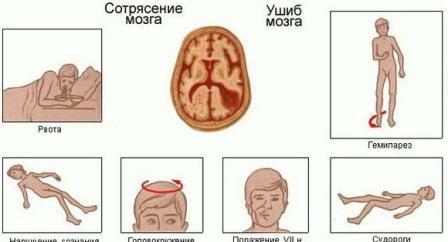
- head injury;
- attacks of numbness of fingers;
- high blood cholesterol;
- noise in the head, ears;
- presence of symptoms of developing acute cerebrovascular accident;
- cervical osteochondrosis.
How to conduct the study
On the eve of the survey is not recommended to take caffeine-containing drugs, drink alcohol or smoke. Also, do not perform the procedure after intravenous injections, as this may affect the result. Excluded are any drugs that affect blood flow and blood pressure, except when taking these medications is vital.
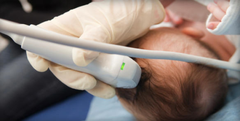
 The patient is positioned on the couch with his head tilted back and set aside in the opposite direction to the examination. The ultrasonic probe is mounted on the neck first from one side. Next, the vascular bundles of the neck on the other side are evaluated. The duration of the study is individual, but on average it lasts 10-15 minutes.
Ultrasound examination of the vessels of the head
Duplex scanning of the vessels of the neck and brain visualizes the vessels not only located on the neck, but also the blood supply to the brain, determining the blood flow through the intracranial vessels. Since bone tissue( skull) serves as an obstacle to the penetration of ultrasonic waves, so-called acoustic windows are used to perform transcranial dopplerography: the temporal region, the eye orbit, the area of the occipital bone and the vertebra.
Duplex vascular examination is performed with:
- chronic head bolus;
- of neurocirculatory dystonia;
- blood flow disturbance;
- dizziness and tinnitus;
- recurrent syncope;
- suspected abnormally located vessels of the brain;
- of discirculatory encephalopathy;
- assessment of the degree of atherosclerotic damage to the head vessels;
- TBM.
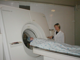
 What diseases are revealed in the tomography of the brain: who is prescribed the procedure, the risks of conducting a survey.
Find out how to conduct the EEG of the brain and to whom the examination is shown.
How is a duplex study of the vessels of the head and neck
? On the eve of the study, vascular drugs that affect the results of the procedure, alcohol and nicotine are excluded. Examination spend lying on a couch. The ultrasonic sensor is installed on the back of the head, in the area of the eye socket and temple. During the diagnostic event, it will be necessary to turn your head to the side, breathe quickly or hold your breath at the doctor's request. The duration of the procedure is no more than 15 minutes.
It is possible to evaluate the results according to the following indices: pulsation and resistive indexes, linear velocity of blood flow and its character, vessel diameter and percentage of occlusion by thrombus, systolic-diastolic ratio.
Ultrasound duplex scanning of the vessels of the head and neck is a modern diagnostic method that allows not only to establish an accurate diagnosis, but also to evaluate the effectiveness of treatment. The examination is appointed by the attending physician. After receiving the results, he determines the further direction of therapy and selects medications taking into account the features of the course of the disease.
write the question in the form below: