All About MRI of the Brain
Contents:
- What is an MRI?
- When is the procedure shown?
- Head injuries
- Tumors
- Inflammation
- Multiple sclerosis, neurodegenerative diseases
- Advantages and disadvantages MRI
- Conclusion
 Magnetic resonance imaging of the brain is a procedure that allows diagnosing tumors, cysts, vascular pathologies, sclerotic changes, inflammation. Why do MRI of the brain, how many times can I do, are there any shortcomings and contraindications to the implementation? The answers to these and other questions are the subject of this article.
What is an MRI?
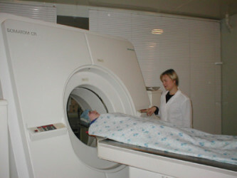
This study of the soft tissues of the head with the help of a magnetic field, the radiation of which is reflected by tissues and fixed by scanners that give a signal to the computer. The patient is laid on the couch, fixed with straps, pushed into a special tube, which is affected by radio frequencies and magnetic field.
 All about the methods of diagnosis of the vessels of the head and neck: indications for the study of the vessels of the head and neck.
Find out what is CT scan and what are the risks of the procedure.
Tomographic images are layered sections of organs. If necessary, use a special contrast agent. Information obtained by this method can be recorded on information carriers and images.
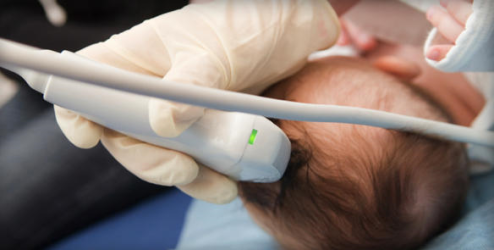
In children, MRI can be performed under anesthesia or sedatives if necessary, if the child can not lie still.
When is the procedure shown?
Magnetic resonance imaging of the head is an expensive but effective study that can reveal a variety of pathologies of soft tissues. Why do MRI of the brain? This diagnostic manipulation can detect the following pathologies:
- Tumors of the head.
- Inflammatory processes in bone sinuses, meninges, its cavities.
- Amyloidosis of neural tissue.
- Brain formation injuries.
- Internal bleeding.
- Neurodegenerative diseases( Alzheimer's, Pick's disease).
- Foci of multiple sclerosis.
- Epileptogenic foci.
- Ventricular expansion.
Head injuries
To make an MRI of the brain is recommended in cases of craniocerebral trauma( TBI) to assess soft tissue damage, to detect bruising in the presence of bleeding. The wedging of the trunk due to the formation of post-traumatic hematoma is a dangerous injury that can lead to death.
The magnetic field created by the scanner, and the scanner that catches the reflected radiation, allow us to see ruptures of vessels, soft and hard shell, venous sinuses. Perform magnetic resonance imaging for signs of TBI, such as unconsciousness, amnesia, vomiting. Diagnosis is shown for bleeding from the ears and nose, as well as the flow of a clear liquid from them - the liquor. It complements the X-ray examination, which allows you to assess the damage to bone tissue.
Tumors
Neoplasms of the head can lead to symptoms such as fainting, dizziness, pain, endocrine disruption( with pituitary tumors).Also, visual, hearing, and balance disorders are possible. Tumors pose a health threat, so they should be detected as soon as possible in order to prescribe treatment or surgical removal.
Inflammation of the
Infectious and inflammatory processes of the brain and its membranes can lead to changes in reflexes, increase the tone of individual muscles, epileptiform seizures. With these symptoms, a magnetic resonance study is assigned. Diagnosis is also necessary in inflammatory processes in the mastoid process, the paranasal sinuses: frontitis, sinusitis, sphenoiditis, etmoiditis.
Multiple sclerosis, neurodegenerative diseases
With MS signs such as vision impairment, eye flies, hearing loss, numbness, tingling of parts of the body or their paralysis, urinary incontinence, an MRI study should be done.
 Senile dementia in Alzheimer's disease is characterized by absent-mindedness, memory impairment, loss of ability to monitor oneself. Amyloidosis is a disease in which a protein similar to starch accumulates in the nervous tissue, disrupting its functioning. Symptoms are similar to senile dementia( dementia).
Advantages and disadvantages of MRI
Pros:
- Relative safety for adults, children, pregnant women after 12 weeks, compared with computed tomography, since X-rays are not used.
- Reliability and informativeness. The study is the gold standard in the diagnosis of all diseases of the central nervous system.
- Non-invasive method.
Cons:
- The cost of the study is from 3000 to 8000 rubles.
- Requires a lot of time( from 20 minutes to an hour) and immobility from the patient.
- It is impossible to do if there are metal objects in the body: plates replacing the bones of the skull, bullets, pacemaker.
- Noise during the research.
- Contrast impact on bone marrow hematopoiesis, kidney( Gadolinium).
- The effect of the magnetic field on the circulatory system, since the blood contains iron.
 All about EEG of the brain: symptoms, preparation for the procedure, results.
Find out how to carry out the brain's CNS and the indications for the study.
Assignment, a variety of methods of tomography and indications for its conduct.
Contraindications:
- Pregnancy up to 12 weeks.
- Alcohol or narcotic intoxication.
- Heavy claustrophobia.
- Presence of tattoos with iron, metal implants: artificial heart valves, insulin pumps.
- The presence of electronic devices in the body( pacemaker).
- For contrasting - a violation of the formation of red blood cells.
Conclusion
Where to make an MRI of the brain? The study is conducted in polyclinics, hospitals and diagnostic centers. Is it scary to do an MRI of the brain? It depends on the state of mind. In case of fear of closed spaces( claustrophobia) or other neuroses, a patient may panic.
How often can I do an MRI of the brain? Such a tomography is relatively harmless, therefore it can be performed with almost no restrictions. Magnetic resonance imaging is a reliable method for examining the soft tissues of the head. Therefore, she is preferred to computed tomography. In addition to this examination for the purpose of differential diagnosis, patients are prescribed electroencephalography, as well as rheoencephalography.
For more information, watch a video of how to do MRI of the brain to children.
write the question in the form below: