Superficial caries
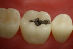
 Surface caries is a long-lasting ailment of solid tooth structures, mediated by a variety of factors and is indicated in the enamel, before contact with the dentin defect. Surface caries on parts of a white or pigmented spot develops with continued processes of decreasing the content of inorganic substance in dense structures of the tooth( the mass of the mineral substance decreases).Visually, the presence of a cavity on various facets of the tooth is often indicated, often where it is more difficult to purify( fissures, interdental spaces).It is determined in persons of any age.
Very often, superficial caries in children is indicated in correlation with a lack of mineralization of dense structures of the tooth. Therapy is performed with preparation, treatment of antiseptics, setting of a seal. Choose the substance for the seals in correlation with years and well-being. The value for surface caries and therapy for replenishing the inorganic substance of the tooth.
Causes of superficial caries
Superficial caries continues the destructive process that occurs during caries during the stain, under the action of microbial constituents in dental plaque, food particles, and lowering of the alkaline balance. Enamel is a solid structure of the tooth, the composition is represented by organic and mineral substance. Moreover, the percentage prevails towards mineral content. Under the action of unpleasant aspects, dissolution occurs at the points with the antigen of the mineral, thereby causing the birth of the defect. If the process is ignored, destruction occurs with the formation of voids, forming a superficial caries. The edges of the tooth, on the sides of the defect in the surface caries become uneven, rough at the touch( the process of dissolution of the mineral content of the enamel passes unevenly and into unequal penetration).
It becomes clear that the starting value of caries at the stain and surface caries is due to unsatisfactory cleaning of the mouth and excessive consumption of carbohydrates. With irregular cleaning on the faces of the teeth, the accumulation of food particles is designated, causing the formation of a film that turns into plaque. Inside this formation, microorganisms that function well in an acidic environment are active. Gradually, the plaque hardens( minerals use from saliva, food), and inside the spread of microbes continue, generating organic acids, causing the dissolution of minerals. Soon there is a defect.
Symptoms of superficial caries
Surface dental caries of the symptomatic manifestations is quite similar to the rest of carious lesions, especially with medium caries. The patient marks formation on the brink of a tooth, with the probability of visualization. It is colored: light, or dark( which corresponds to the course of the process of superficial caries: the acute stage is transient and corresponds to a light color, the chronic is inherent in a prolonged direction and therefore the defect will be dark).The patient basically notes the pain instantly conditioned by the use of sweet, sour, salty. Discomfort may be indicated for the use of hot and cold liquids.
Discomfort in the superficial caries on the background of the designation of the approach to the outer edge of the tooth vascular-nerve weave at the cavern. The pain will appear and with excessive toothbrush toothbrush region of the neck of the tooth when marking the cavity( the thickness of the enamel is low, and with superficial caries is significantly reduced).
If the patient tries to investigate this defect with his own tongue, then when touching with superficial caries it will indicate roughness. In general, there is no transformation with superficial caries. When docking and self-exerting tension on the causal tooth and its antagonist, nothing is indicated. When the patient visualizes the gums and oral mucosa, there are usually no changes either. However, if the superficial caries is identified between the teeth, the patient determines the jammed food and slight pain when pressing on the tops of the gingival papilla( qualitative cleaning of the faces was not performed, and the pressure of excessive contents was carried out).
Diagnosis of superficial caries
Surface caries is diagnosed by collecting a general picture based on the results of the survey, noted changes in the mouth during visualization, addition of minor research methods. The patient survey provides an opportunity to collect a preliminary understanding of the process characteristics and the approximate depth of the lesion. The patient notes a short period of pain from derivatives of chemical nature( sour, sweet, salty).To note the patient at a superficial caries from reception of drinking of different temperatures the pain can( more is shown at a designation of a carious cavity about a neck of a tooth).Sometimes patients with superficial caries highlight an increase in sensitivity and hygiene of the dentition( especially when using a hard brush) against this background. When examining the cavity in areas accessible to visual inspection, the patient can indicate a change in the color of the tooth and a defect, when the tongue is placed along the side of the tooth, roughness is established. Examination of the tooth gives already more pronounced symptoms for diagnosis. Visually, there is a defect of matte color in the projection of the crown.
The clinic of superficial caries is based on sensing data, water samples, percussion, palpation of the transitional fold in the apex of the causative tooth, examination of the mucosa. Probing will help to clarify the carious defect, assess the depth, the presence of pain on the bottom of the cavity, whether there is a connection with the cavity of the tooth. There is also a condition of the seals adhering to the tooth. Produce this technique through a dental probe. During the examination, the doctor determines the following indices: the intensity of the lesion, PEC, prevalence, increase in morbidity( regular examination is necessary).Also, a "Caries Detector" is used by the doctor to detect a non-vital layer of the carious cavern( apply a swab with the substance, into the cavity of the surface caries and wait a quarter of a minute, the non-viable layer will become colored).
The clinic of superficial caries during probing will be marked by a tool jam, the presence of uneven edges, and the pain of the bottom of the cavity may not be noted. A sample of water in a carious cavern can give a short-term pain, quickly arising( the vascular-plexus of the tooth is still alive).Percussion and palpation of the transitional fold will not show changes( hence, there are no inflammations of the sites in the periapical tissues).Mucous membrane without deviations. If superficial caries of teeth is noted at the points of contact of two teeth, then one can note food with a plaque in the cavity, swelling of the gingival papillae( the contents of the cavity exerts pressure on the gum, triggering inflammatory phenomena in it).Additional methods: EOM, transillumination method. Conduction of EOM( electrodontometry: study of the sensitivity threshold of pulp tissue to electric current) will not yield informative data, since its indications will be noted within the limits of the norm, 2-6 μA.The transillumination method will reflect when using a light guide made of organic glass, connected to a dental mirror in a dark room, areas of darkening of the teeth that belong to the surface caries.
Treatment of superficial caries
Therapy of superficial caries is carried out in correlation from the depth of the enamel defect. In case of a violation in the external department, a defective facet is grinded and surface preparations are applied to repair the inorganic tooth matrix( Gluftred).If defect deep structures in the surface caries, with the preparation must be treated. Ideally, the tooth is separated from a number of existing structures, with the support of a cofferdam, ruberdam. Manipulation in the mouth is recommended to be performed with air-water intake. At the same time, the aspiration system in the therapy of superficial caries is simultaneously recommended, since this minimizes the time for squeezing the patient's fluid from the mouth. The cooling of the tooth is the prevention of overheating of the pulp tissue. Since the speed of rotation of the working parts on the tips is high, and the frictional force about the tooth increases, these processes are combined with the release of a significant amount of heat. With excessive overheating, the vascular-neural interlacing dies, and the process from the carious course can be transformed into periodontitis. The preparation of tissues is precisely necessary in places of high risk of caries spread: deep fissures, interdental contacts.
Before the opening of the carious cavern, the teeth are cleaned from the plaque and stones. Initially, the removal of hard stone by ultrasonic scaler, combining with the tools for removing the calculus by hand( excavator, periodontal hooks, if necessary).When the patient notes uncomfortable feelings, local anesthesia is performed: application( Lidocsor-gel or spray), infiltration( Articaine, Ubistesin, in the interdental papillae in a small lobe).Then, a soft plaque is taken with the Air-flow apparatus( an air-water substance with a specific powder based on sodium bicarbonate is applied to the tooth).Further, special brushes are used for the micromotor tip, in conjunction with the paste for hygiene( Prophylactic Paste( firm PD)).And finally, the dissection of the tooth begins.
In the identification of pain for intervention in superficial caries, anesthesia is performed. Perform the preparation with burs on the turbine tip. Antiseptic wiping( Chlorhexidine bigluconate 0.05%).Further therapy is due to the type of filling substance used. When setting a light seal, etching is used. Apply a dressing with a content of 30-40% orthophosphoric acid( Travex 37), gel holding for 15-30 seconds. Flushing with water from the aspiration system by adding, drying the cavity. Then use an adhesive( adhesive for the connection) of different generations. Possible application for surface caries and self-etching adhesives. Then the stage of using acid is not needed. After application of the adhesive, blowing is performed, using a small feeding force to the corresponding section of the air-water gun. They are illuminated with a photopolymerization lamp for a period corresponding to the instructions of the preparation. Apply a gasket with fluoride content( Vitrebond( prevention of recurrence)).If, according to the manufacturer's instructions, it is necessary to flash, then it is carried out. The restoration is performed with a light composite: layer filling of the cavity, with a single application and distribution of material 1-2 mm thick. After each stage of the attachment, a flash is made. After finishing( removing surplus fillings, roughness( using burs, abrasive discs, brushes with pastes)), apply fluoralk( preventive maintenance of relapse), or perform rebonding( use of an adhesive - preferably self-etching).

surface caries: photos of the child
Surface caries in children, preferably pre-school and early school life intervals, is preferably treated with fluorine-containing cements( glass ionomer): after dissection and antiseptic treatment, the cement is applied containing fluorine( Kemphil).It is also possible to use light cements( Vitrimer).Use according to the instructions. After the setting, the finishing is done, taking into account the material( the cement should harden and it takes some time, sometimes the next step is performed in a day).After finishing, fillings are filled with fluorinated varnishes. Preference in the therapy of superficial caries is given to the cement, because the use of them minimizes the cost of setting the seal, which is of great importance for the treatment of children's teeth.


