Biopsy: how is biopsy done?
Biopsy studies are often used as a method of modern diagnostics in medicine.This study was based on the lifetime taking of a biomaterial( tissue) in a patient for the purpose of microscopic examination.
The research process itself involves taking the material, fixing it securely, transporting it to the laboratory, where it is invariably processed, then the sections are made and painted.And only after all these procedures you can start a microscopic study, which will help to diagnose.
A biopsy is advisable in the case when other methods are poorly informative in terms of diagnosing.In this case, a biopsy is always prescribed for suspected malignant tumors.
Table of contents: Biopsy types Biopsy methods Biopsy methods Biopsy biopsy methodsBiopsy types
Biopsy happens:
- Partial when a piece of tissue is taken from the education center for research.It is also called incisional biopsy.

- Total, in which the examined pathological focus is completely removed.This procedure is called excision biopsy.
These two types of biopsies are used by surgeons in the process of interventions and they are performed exclusively in the operating room.
Biopsy Techniques
The most commonly used biopsy techniques are:
- Surgical open biopsy, which is assigned during surgery.
- Closed biopsy
Closed biopsy is divided into:
- trepan biopsy( requires special equipment and a thick needle);
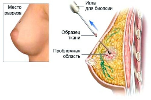
- fine-needle biopsy( performed with a conventional thin needle);
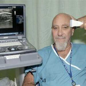
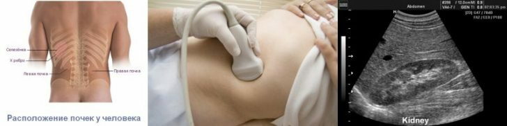
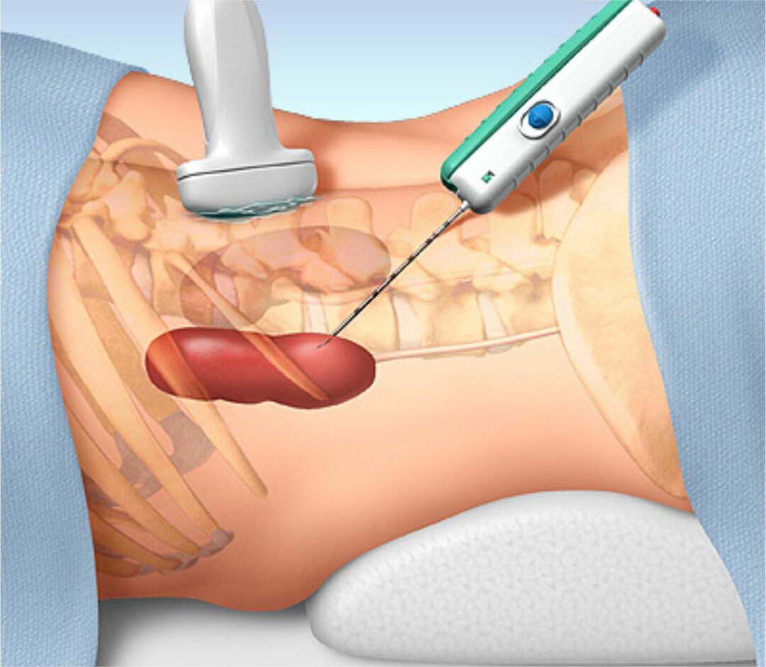
- biopsy under the supervision of ultrasound or X-ray;
- biopsy in the process of fibrogastroscopy;
- endoscopic biopsy performed during gastroscopy;
- biopsy with fibrocolonoscopy;
- biopsy during bronchoscopy.
The most commonly used in practice is thin-needle biopsy. Indications for it are the following conditions:
- reactive lymphadenopathy;
- pathology of the mediastinum, chest wall;
- diagnosis of the liver in the presence of her patprocesses of focal and diffuse nature;
- neoplasm of unknown origin in the adrenal gland;
- pathology of soft tissues;
- abscess of the spleen and its focal primary lesions;
- suspected of a kidney tumor;
- Malignant lymphoma;
- pseudocystic and cystic neoplasms;
- pulmonary disease;
- pancreatic cancer;
- cyst thyroid;
- pathology of retroperitoneal space;
- ascites;
- pathology of the gastrointestinal tract;
- metastases in the lymphatic system.
This biopsy is performed in an outpatient setting.
There are also a number of contraindications to it:
- patient's refusal in writing;
- severe blood coagulation system pathologies;
- is a tumor with suspected melanoma;
- having the ability to perform a more informative but non-invasive study;
- threat of miscarriage.
Biological material research methods for biopsy

There are two types of such methods:
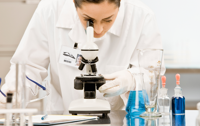
- Cytological examination.It involves the study of cells taken from a biopsy from the surface of the tumor.It is a technology of cytomorphological diagnostics, due to which the character of the neoplasm is determined( precancerous, malignant, reactive, benign, inflammatory).The preparation of the preparation is as follows: the cut of the surgical material or biopsy touches the glass on which the imprint remains( a thin smear), it is stained and studied under a microscope.
- Histological examination.It is carried out in a planned and urgent manner.A planned study of cells in biopsy involves placing the tissues in a special solution, and then - in paraffin, then perform slices and staining.Such a survey takes about a week's time.Urgent study of tissues is carried out by freezing tissues.A microtome( knife) is cut, and the staining is carried out by the doctor under a microscope.The duration of such diagnostics is up to 40 minutes.Usually, urgent research is used during the operation to determine its volume and the nature of the tumor.
Decoding of biopsy results
The norm is the absence of cell changes in the study of biopsy material.
Viktorova Julia, obstetrician-gynecologist