Types of microcephaly and the causes of the pathology
Contents:
- Prognostic factors of the disease
- Distinctive features of microcephaly
- Diagnosis of microcephaly before and after birth
- Principles of treatment and prognosis
 Microcephaly belongs to congenital malformations, the causes of which are associated with many factors. The mass of the brain in such children is much less than the norm, respectively, the skull is formed of reduced sizes, and the baby lags behind in mental development. Let's consider why the disease occurs, how are the predictions for the baby and his parents manifested?
Provoking disease factors
Distinguish primary microcephaly( true), caused by genetic factors and secondary, due to a mother's disease during pregnancy. Genetically determined microcephaly can proceed as an independent form:
- Pein syndrome - mental retardation, leg muscle cramps, heart defects that occur exclusively in boys( recessive disease linked to the X chromosome);
- Giacomini syndrome - the presence of convulsions, paralysis and mental development defects in members of the same family.
Also microcephaly is one of the manifestations of chromosomal defects( Down syndrome, Patau syndrome, Edwards syndrome and others).
The development of the secondary form of the disease is associated with the damaging effect of certain factors on the baby's body during the period of the bookmarking, the formation of the brain. Let's present the most common reasons:
- Infectious diseases of the mother. To dangerous factors include toxoplasmosis, infection occurs when contacting domestic and domestic cats, when consuming insufficiently cooked or roasted meat. Also, infection with measles, rubella, cytomegalovirus viruses in the first weeks of pregnancy leads to the death of the fetus or pathology of the nervous system. Dangerous viral diseases include Zika's fever.
- The effects of alcohol, nicotine, narcotic, toxic substances - penetrate the placenta and mutilate or kill the fetus.
- Ionizing radiation - radiation, a long stay near the TV, various computer equipment increases the risks of congenital brain pathology.
- Diseases of the mother, trauma, causing hypoxia or mechanical damage to the baby.
- Fasting or insufficient intake of essential nutrients( proteins, lipids, carbohydrates, minerals and vitamins) for the formation of fetal organs.
Distinctive features of microcephaly
The presence of pathogens is determined at the first examination of a newborn, reveals the basic symptoms of microcephaly:
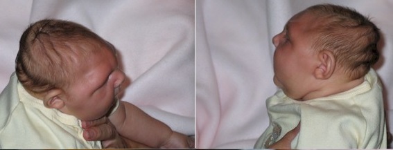
- The child has features - mainly the facial skull is developed, its sizes exceed the size of the brain part. The face of the baby grows with age, the size of the head changes slightly.
- Weight, the size of the brain abruptly( on average half as much) differ from normal indicators. The circumference of the newborn's head is 25-27 cm, with a norm of 35-37 cm.
- Appearance - the forehead of the patient is high and steep, the superciliary arches protrude, against the background of the small head, the ears seem quite large. Often there is a hare lip, strabismus or wolf mouth.
Important! The baby has a closed fontanel or it closes in the first month of life. Earlier ossification of the fontanelle serves as a differential sign of microcephaly in newborns.
Emotional sphere and mental abilities depend on the severity of the disease, the kid can be hyperactive or lethargic, serving himself and studying in school( with moderate backwardness) or utters individual sounds and needs constant care( with oligophrenia).The disease can be combined with epilepsy or cerebral palsy, accompanied by cramps, paralysis or a violation of muscle tone.
Diagnosis of microcephaly before and after birth
 There are a number of methods for diagnosing fetal microcephaly during pregnancy. The basic method is ultrasound with the most accurate data after 27 weeks, with a shorter period of time, there are false positive or negative results. The study of amniotic fluid refers to more accurate, but invasive methods. In some cases, it can provoke premature birth.
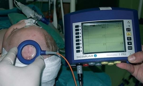
Microcephaly of the brain in small and older children is determined with the help of CT, MRI, neurosonography. Detecting the structure of the cerebral hemispheres and frontal lobes in particular, cysts are formed in the brain tissues, the number of convolutions is reduced or they are smoothened and absent.
Principles of treatment and prognosis
Treatment of microcephaly includes several tasks:
- specific drug therapy in combating the cause of the disease;
- symptomatic treatment - reduction of intracranial pressure, anticonvulsant therapy, improvement of mental activity;
- vitamin and mineral complexes;
- physiotherapy, massage, swimming.
The consequences of microcephaly depend on the degree of brain damage. If vital centers are hurt, the baby dies on the first day after birth. The presence of manifestations of epilepsy or cerebral palsy increases the risk of early death of the child. Primary microcephaly with chromosomal abnormalities has an unfavorable prognosis. Children have problems with immunity, and they can die from infectious diseases before the age of 15.
However, with insignificant mental retardation, proper care and application of pedagogical and developmental methods, children with microcephaly disease practically catch up with their peers, study and lead an active lifestyle.
write the question in the form below: