Fallato heart tetrad Phallo
 In modern medicine, the disease of the tetralogy of Fallot is not uncommon. In 7% of all cases of heart disease there is a tetralogy of Fallot. The so-called "blue heart" was identified by the French pathologist Phallo, who gave the definition of the disease.
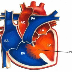
The tetralogy of Fallot is a type of heart disease that has four basic anatomical symptoms:
- Defect of interventricular septum;
- Incorrect aorta or aortic dextrase;
- Obstruction of the flow of blood from the right ventricle( stenosis of the pulmonary artery);
- Hypertrophy of the right ventricle;In addition to these anomalies, coronary artery anomalies, the right aortic arch, the presence of aortolateral collateral vessels, pulmonary artery stenosis, an open arterial duct, regurgitation on the aortic valve, and many others can also be observed.
Reasons for the development of tetralogy of Fallot
The most common cause of the disease are genetic abnormalities. These include: Down syndrome, Patau, DiGeorge syndrome( deletion of the long arm).Other common causes are endocrine diseases, in particular diabetes maternal, vitamin A intake in large doses before pregnancy and already during pregnancy. The intake of alcoholic beverages, the use of narcotic and psychotropic substances, which can also cause the development of heart defects in the tetralogy of Fallot.
Among the reasons can also be identified physical factors, such as X-ray radiation, ionizing radiation.
Chemical factors - nitrites, phenols, drugs, including antibiotics, antitumor drugs and others.
Biological factors - viruses of infectious diseases, reduced immunity, broken amino acid metabolism, systemic lupus erythematosus and others.
Normally, circulating blood in the body is enriched with oxygen in the lungs. But with Fallot tetrada disease, the narrowed right ventricular duct interferes with the enrichment of blood with oxygen in the lungs and thus the blood acquires a "blue" hue.
Symptoms of the disease
In newborns, the symptoms of the disease may not appear immediately. But after a few days, the work of the heart can be significantly disrupted and the first symptoms of the disease appear.
One of the symptoms is cyanosis or cyanosis of the baby's skin. So cyanosis can appear when feeding or in a calm state. Most often, the blue shade acquires auricles and lips. Then cyanosis can acquire a more pronounced shade.
There may be shortness of breath, which is worse with force. Sometimes a patient can not even get out of bed. In such cases, immediate rest and peace is needed.
Over time, the fingernails and fingers of the patient can take the form of clock hands and drum sticks, respectively. This is due to impaired blood circulation.
Due to the degree of development of the disease, several phases of the tetralogy of Fallot are distinguished. Each phase corresponds to the symptoms of the disease and age. There are three phases:
- I phase - relative well-being. Develops at the age of up to 6 months. During this period, there is no noticeable lag in the development of the child.
- II phase - phase of dyspnea and cyanotic seizures. It develops at the age of 6 months to 24 months. There are attacks of cyanosis, dyspnea, which lead to cerebral complications, delayed development. This is just the case when blood is not rich in oxygen, which leads to brain starvation. During this period, systolic murmur subsides, the child is frightened, especially cyanosis, the child loses consciousness, and then convulsions begin. After an attack the child is sluggish and motionless. In some cases, seizures lead to death.
- Phase III - transitional, in which the symptoms begin to resemble the symptoms of an adult.
Diagnosis of the disease
When diagnosing the tetralogy of Fallot, first of all, an external examination of the patient is carried out. The development, growth of the child, and the transferred diseases are studied. Attention is drawn to the condition of the skin and its color. The skin is examined for the presence of cyanosis, the finger phalanges and nail plates are inspected. Then, the heart sounds are heard. Particular attention is paid to the examination of the chest, in which the "heart hump" can develop. Between the 2nd and 3rd ribs, systolic jitter can occur.
It is not superfluous to carry out chest X-ray. As a rule, cardiomegaly is found( enlargement of the heart), which manifests itself in a specific increase in the heart in the form of a "shoe", "boot" or "sabot", in which the heart's waist increases and the apex of the heart rises.
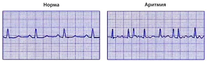
When performing an ECG, it is possible to detect a deviation of the electrical axis from the norm from about 120 to 180 degrees to the right side.
When examining the heart with ultrasound, you can find all the symptoms of the tetralogy of Fallot: hypertrophy of the right ventricle, obstructive movement of blood from the right ventricle, incorrect location of the aorta and defect of the interventricular septum.
It is also possible to probe the heart cavities, in which the pathology of the pulmonary artery and the presence of collateral blood flow can be detected.
Angiography is a mandatory method for diagnosing heart disease with contrast agents. This method allows you to determine the condition of the heart and the forecast for the operation.
Treatment of tetralogy of Fallot
People suffering from Fallot tetrad disease in any case will be subjected to prompt intervention.
In the acute course of the disease, surgical intervention is given immediately.
If the disease is detected in a child under the age of three, then palliative operations are appointed, consisting in increasing the flow of blood in a small circle of circulation. For palliative or bypass operations include anastomosis Bleloka-Taussig, central aorto-pulmonary anastomosis, valvuloplasty, etc. By
dyspnea and cyanotic attacks assigned drug therapy:. Intravenous administration of sodium bicarbonate, aminophylline, glucose reopoliglyukina. Also prescribed are inhalation with oxygen.
Postoperative complications
These complications include: pulmonary hypertension, arrhythmia, right ventricular aneurysm, congestive heart failure, infective endocarditis, and others.
Mortality during the operation does not exceed 5%.After the operation, the quality of life of the patient is much improved: the cyanosis of the skin disappears, the attacks of dyspnea decrease, the results of the tests improve. Patients with Falla tetrad should always be under the supervision of a doctor.
Prevention of the disease
- Timely account of the state of the body in a doctor;
- Compliance with proper nutrition and day regimen;
- Elimination of the influence of harmful environmental factors;
- Exclusion of drugs, drugs;
- During the period of exacerbation compliance with bed rest and rest;
- During pregnancy, systematic observation by a doctor;
- Correction of the state of the organism depending on the concomitant diseases.