ECG: what it is, how to make a cardiogram of the heart correctly and what does it show
Content:
- Technique
- Views
- Training
- Norm indications
More than a century has passed since scientists discovered the ability of the heart to produce electrical impulses in small doses.
This discovery marked the beginning of the science of electrocardiology, an integral part of which is electrocardiography. This section examines the electric current that occurs in the heart, or affects it from the outside.
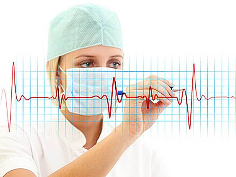
Electrocardiography is able to register electrical potentials arising from relaxation and contraction of the myocardium in a certain time interval.
These impulses spread throughout the body and reach the skin.
A special device - an electrocardiograph - fixes these potentials and gives the result in the form of a graphic image, called an electrocardiogram. It can be printed on paper or displayed on the monitor screen.
Electrocardiography can serve a variety of purposes:
- To assess the effectiveness of cardiac drugs, pacemaker and other methods of therapy.
- To identify and trace the dynamics of the development of heart diseases such as arrhythmias, disorders intracardiac conduction (blockade) and metabolism necessary for the work of the heart (potassium, magnesium, calcium). Here you can also determine myocardial damage, the physical condition of the organ, acute cardiac pathologies and non-cardiac diseases (for example, pulmonary embolism).
ECG is a very simple procedure that has practically no contraindications. It is allowed to do it for women during pregnancy and even for newborns upon discharge from the hospital. In emergency cases, the cardiogram is removed by an ambulance paramedic in a special car, at the patient's home and even on the street.
Most often, the procedure is carried out in district clinics, hospitals, specialized clinics, sanatoriums. This takes no more than 10 minutes and does not cause absolutely any discomfort to the subject.
However, with all the positive aspects, electrocardiography also has disadvantages. Here, the short duration of the procedure is most often noted.
The principle of operation of any electrocardiograph is based on the propagation of cardiac impulses. They are able to move by decreasing the polarization of the cell electrodes. At rest, the surfaces of all muscle cells in the heart are positively charged.
At such a moment, there is no potential difference and, accordingly, it is impossible to register the electric field.
Electrical impulses in the heart usually originate in the sinoatrial (sinus) node.
It is located near the superior vena cava in the right atrium. The node is a specialized cell with the ability to automatically generate electrical impulses. The latter spread from the sinoatrial node, first to the right, then to the left atrium.
The result of the spread of electrical signals through the atria and ventricles is their contraction. The result is the flow of blood to the lungs and to the circulatory system.
Cardiogram of the heart: registration technique and scope
The recording of the potential difference between two points of the heart's electric field with an electrocardiograph is called a lead.
When recording a cardiogram of the heart, standard leads are recorded from two limbs by alternating paired connection of electrodes. Three standard positions form a triangular shape (Einthoven's triangle).
Recording of a cardiogram of the heart is carried out in a calm state of the patient. In some cases, a specialist records an ECG on inspiration, asking the patient to take a deep breath.

At analysis of ECG results the cardiologist must have the necessary knowledge and skills to decipher the graphic image.
Electrocardiography is prescribed not only for existing heart diseases or suspicion of them. The doctor may recommend doing an ECG as a preventive measure, as well as during medical examination and annual medical examination.
If there is no suspicion of abnormalities, a cardiogram of the heart is done upon receipt of a medical record for a job. For children, an EKG is done upon admission to the kindergarten, and according to the new rules, it is obliged to provide it to the head of the sports section, the guys involved in it. In addition, ECGs are often done to pregnant women before childbirth. Patients with diabetes mellitus must be examined without fail, even in the absence of indications.
The referral for the study is given by the attending physician or cardiologist. Indications for an urgent procedure are pain in the region of the heart, fainting, dizziness, hypertension, swelling of the legs, weakness in the joints.
Electrocardiography: types of diagnostics
 The first device capable of recording a high-quality ECG was a string galvanometer designed by V. Einthoven. Its basis was a very thin thread, which was in a magnetic field under a certain voltage. He created a new direction in the physiology of blood circulation - the electrophysiology of the heart.
The first such technique was very bulky and weighed 270 kg.
IN. Einthoven marked the main waves, intervals and segments of the ECG, and also calculated their time intervals. He also proposed a system for placing electrodes on the surface of the patient's body. These data are used by cardiologists to this day.
Prongs are the ups and downs in a graphical image. A segment in electrocardiography is a section of a straight line between two teeth. An electrocardiogram is able to show violations of the heart in the early stages, as well as consider the possibilities for the development of serious pathologies.
However, the ECG does not always accurately determine the presence of the disease. For example, a violation of the rhythm of the heart (arrhythmia) at the time of the study at rest may "hide" and not manifest itself.
Therefore, the specialist chooses a different examination method, there are only a few of them:
- At rest Is the most commonly used standard method. The patient lies relaxed on the couch.
- With load - during this procedure, the doctor will first take the readings of the electrocardiograph, then ask the patient perform a simple physical exercise (bends, squats), and then re-examine with the help apparatus. In addition, it is possible to use other methods - bicycle ergometry and treadmill test. In the first case, a bicycle ergometer (an apparatus similar to an exercise bike with varying pedal resistance) is used, in the second, a treadmill (a moving track). For each type of examination, electrodes connected to a computer are applied to the patient's body. During the procedure, the doctor monitors and analyzes the indications.
- Daily (Holter) monitoring - this method is the longest in time. When using it, adhesive electrodes are attached to the subject's body. They connect to a device that is attached to a belt or worn over the shoulder on a belt. It weighs no more than half a kilogram, so it does not cause any particular inconvenience.
The patient should keep a diary, which indicates information about changes in physical activity, emotional overload, time of taking medication, sleep and wakefulness. Here he describes pain in the heart and a feeling of discomfort that may arise during certain activities.
There are two options for Holter monitoring: full-scale and fragmentary.
The first continues continuously for
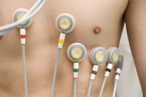
Fragmented monitoring can take longer. They resort to it only when cardiac malfunctions occur infrequently. In this case, electrocardiography is performed using a special device.
To register deviations, the subject turns on the ECG recording button when pain appears. The apparatus for such research is rather miniaturized: it can be a pocket version or a device in the form of a wristwatch.
A transesophageal ECG is recommended to be performed with low information content of standard methods.
A sterile electrode is inserted into the esophagus. This is usually done through the nasopharynx, less often through the mouth. In this case, the patient must make swallowing movements. But do not be alarmed - the probe for transesophageal electrophysiological examination of the heart (TEPFI) is thin and its introduction usually does not cause difficulties. At the same time, electrodes are attached to the chest to register an electrocardiogram.
The electrode is inserted approximately 40 cm - where the heart is closest to the esophagus. After that, they begin to record a cardiogram, and weak electrical signals to the heart are sent to the probe, forcing it to contract more often.
At the end of the study, the electrode is removed from the esophagus.
In electrocardiography, there are instrumental methods for studying the work of the heart muscle. These include, for example, phonocardiography. In this case, a special microphone captures the sounds made when the heart muscle is excited and relaxed. As a rule, listening is performed by an experienced professional with good hearing, who is able to separate murmurs and heart sounds from pathological sounds.
In the book by VV Murashko "Electrocardiography" there are other methods of conducting research. Its cost is low, but it will be very useful for those who want to master the basics of ECG.
How to do an ECG correctly: preparation and procedure
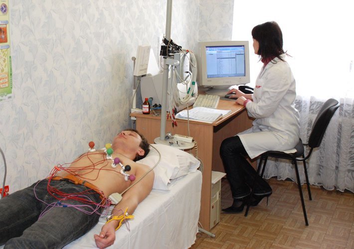 Those who do not know how to do an ECG correctly do not have to worry: electrocardiography does not require special training. However, some nuances still exist. It is advisable to refrain from eating heavy food 2 hours before the procedure.
Also, do not be nervous, play sports, drink energy cocktails or alcohol, as well as strong coffee or tea. Before the examination, women do not need to apply lotion or cream to the body; they should remove any jewelry from the wrists and chest area: bracelets, rings, chains, etc.
The chest electrodes have a special suction bulb, which is sucked to the body due to the created vacuum. The specialist who takes the readings knows perfectly well how to do the ECG correctly, so he is unlikely to be able to confuse the wires connecting the suction cups to the device.
Before starting work, the device must be warmed up (enough
There are no contraindications for conducting an ECG - the study can even be carried out for infants.
In this case, the procedure for removing data from a child is similar to that for an adult. Only the result will be different - for example, babies have a higher heart rate.
Some kids are afraid of all people in white coats, so they can get very anxious before the procedure. Before it starts, parents should relieve the stress of their children - give them their favorite toy, show a funny picture or photo (you can use it on your phone). An older child can be told in advance about the study and in a playful way show how to do the ECG correctly.
The examination procedure may be difficult for individuals with complex chest injuries, high obesity, or excessive hairiness of the chest - in this case, the electrodes will not fit snugly to the skin, and the test result will be distorted. The presence of a pacemaker will also lead to incorrect results.
A transesophageal examination should not be performed in the presence of tumors or other diseases of the esophagus. Exercise ECG is contraindicated in acute infectious diseases, chronic heart failure, coronary heart disease, complex rhythm disturbances, in the acute period of myocardial infarction. Also, you should not do this with an exacerbation of diseases of other body systems - urinary, respiratory, digestive.
A normal cardiogram of a healthy heart and what it looks like
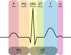 In a healthy adult, a sinus rhythm is considered a normal cardiogram (cardiogram of a healthy heart).
Heart rate (HR) is
PQ interval (period of the excitation wave passing through the atria and atrioventricular node to the ventricular myocardium) -
Changes in rhythm or tonality (arrhythmia, bradycardia, tachycardia) were not found.
Heart palpitations are possible in pregnant women or overly emotional individuals. In elderly patients, on the contrary, there is a slowdown in the rhythm of the heartbeat or morphological pathologies of the myocardium.
Only a specialist with medical education is able to correctly decipher the cardiogram and describe the obtained ECG parameters.
Electrocardiography is capable of diagnosing various diseases of the cardiovascular systems - ischemia, deviations in the development of pathways, heart aneurysm, extrasystole, angina pectoris and many other.
The most serious diagnosis in electrocardiography is myocardial infarction. It is here that you can first detect areas of damaged or dead tissues, determine a specific place (in which wall of the heart) and the depth of the lesion. ECG easily distinguishes the acute phase of a heart attack from old scars and aneurysms.
With a heart attack, the ECG procedure is performed more than once. The first time this happens at the first contact with the patient - at home, in an ambulance or in the emergency room of a hospital. If there are no changes in the graphic image, but if there are symptoms, the procedure is repeated after 6 hours - by this time the symptoms usually appear in full force.
After that, the diagnosis is carried out daily, and upon recovery, every few days. Thus, for the entire period, the patient is examined at least 10 times.
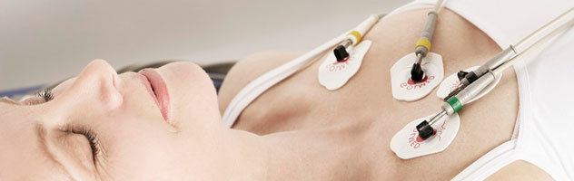 The patient should always remember that only a specialist should be trusted to take care of his or her health. This fully applies to the electrocardiography procedure. You cannot neglect the doctor's prescription and you should not try to decipher the ECG yourself, even if you are sure that the result will be a normal cardiogram.
A cardiogram of a healthy heart, like an ECG with abnormalities, can only be read correctly by a doctor.
Only a person with a medical education is capable of the data obtained as a result of the examination, clinical symptoms and the result of the study, assessing the risk of a critical condition. Otherwise, there is a possibility of underestimating the ECG, which can lead to fatal consequences.
Partial use of site materials (no more than 30% of the content of the article) is allowed only with a hyperlink on www.med88.ru, full use of the article (more than 30% of the article content) is possible only with written permission edition.