Duplex of the vessels of the head and neck: indications for the procedure, the course of the examination and interpretation of the results
 Ultrasound examination is by far the most common non-invasive method diagnostics of various diseases, monitoring the effectiveness of treatment, assessing the condition of the fetus when pregnancy.
However, modern opportunities Ultrasound are not limited to identifying abnormalities in the structure of internal organs. The use of Doppler sonography in combination with "classical" ultrasound allows us to assess the state of the main arteries and veins, the speed of blood flow in them.
This technique is called vascular duplex.
Ultrasound is based on the ability of ultrasonic waves to change physical properties depending on the density of the medium through which they pass. Each type of fabric has an acoustic resistance.
Therefore, reaching the border of a certain organ, blood vessel, hematoma or cavity filled with pus, exudate, ultrasonic waves undergo specific changes.
Some of them are reflected, and some pass further. Thus, a gray-scale image is formed on the screen. Doppler ultrasound is a modern technique of ultrasound examination. This method consists in using the so-called Doppler effect for diagnostics.
Its essence lies in the fact that ultrasonic waves are reflected from moving objects in different ways. Roughly speaking, in the blood these "moving objects" are red bodies, erythrocytes.
Therefore, the use of Doppler ultrasonography makes it possible to assess the blood flow velocity (it is automatically calculated by the device during the procedure) and the degree of vasoconstriction.
This procedure takes place in several stages:
- B-mode, from which any ultrasound examination begins. As a result, the doctor receives a two-dimensional image in grayscale in real time.
- Directly dopplerography. Currently, a color mapping method is used. It can be streaming or energetic. The flow method shows disturbances only in the large main arteries and veins, while the energy more informative, since it allows you to assess the state of the entire vascular bed, regardless of the diameter of the vessel.
Ultrasonic duplex of vessels (USDG) intended for the assessment of disturbances in the blood supply to the brain, neck, heart and lower extremities.
As a preventive measure, such a study is recommended:
- men and women over 40;
- if you are overweight;
- people suffering from diabetes mellitus, thyroid pathologies;
- with a hereditary predisposition to diseases of the cardiovascular system;
- with an increased concentration of cholesterol and low density lipoproteins in the blood.
Duplex scanning of the main arteries of the head, neck and lower extremities is necessary in such cases:
- a previous stroke, heart attack;
- diagnosed heart pathology;
- arrhythmia;
- atherosclerosis;
- suspicion of attached to the vessel wall thrombus;
- preparation for surgery;
- complaints of symptoms of cerebrovascular disorders: memory disorders, hearing disorders, headaches, fainting, dizziness;
- phlebeurysm;
- thrombophlebitis;
- thrombophilia;
- assessment of the consequences of a stroke and the possibility of restoring the patient's health;
- vasculitis.
Also, a duplex of vessels is regularly done to monitor the effectiveness of the prescribed treatment for thrombosis.
Decoding of ultrasound is based on the results of scanning such vessels of the carotid region:
- extracranial on the neck (carotid, vertebral, brachiocephalic arteries, brachial trunk);
- intracranial vessels of the base of the brain (middle, anterior, posterior cerebral, vertebral arteries, venous blood flow);
 With duplex vessels of the lower extremities, the anterior and deep femoral arteries, the anterior and posterior tibial recurrent artery, and the popliteal artery are visualized.
It is difficult to assess the state of the parts of the bloodstream below the popliteal fossa of the leg on Doppler ultrasound, therefore the results of ultrasound require additional confirmation.
The main advantage of the vascular duplex is the absence of contraindications. Study of brachiocephalic and other vessels of the head, lower extremities and heart by ultrasound diagnostics can be performed on a patient in any condition (for example, after paralysis, in coma) without the risk of developing complications.
Also, duplex scanning of the vessels of the head and lower extremities is more common and affordable in terms of price; it does not require hospitalization. The disadvantages of the method include the dependence of the interpretation of the results on the qualifications of the doctor conducting the ultrasound.
Duplex of the vessels of the head and neck: preparation for the procedure, cost, interpretation of results
 In modern medical centers, they can offer a triplex examination of the vessels of the brain and neck. By and large, the procedure does not differ from how the duplex of the vessels of the head and neck is performed.
However, with triplex, the image on the screen of the ultrasound machine is displayed in 3D mode, which gives this technique a great diagnostic value.
No special preparation for ultrasound is required. However, if the patient is taking any medications, it is necessary to consult a doctor before an ultrasound scan. You may need to stop taking medication for a while.
In addition, the doctor who is doing the ultrasound should be informed that the patient is undergoing treatment. You also need to voice the specific names of the drugs.
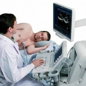
Duplex scanning of the vessels of the brain, carotid, vertebral artery is performed in the supine or sitting position. The sensor is placed on the skin in the projection of the investigated vessels, in the area of the skull bones, which transmit ultrasonic waves as much as possible.
Assessment of the blood flow of the lower extremities begins from the supine position, the sensor is first placed in the area towards the inguinal fold, and then moved in the direction of the vessels. To examine the popliteal artery, the patient is asked to roll over onto his stomach.
The procedure is painless and takes about half an hour. Its cost depends on the region and clinic. On average, the price of ultrasound and duplex of the vessels of the head and neck ranges from 2,500 to 3,000 rubles.
Deciphering the survey results allows you to identify the following deviations:
- determination of pathologies of the vascular bed of the brain before the appearance of the first clinical symptoms of diseases;
- the presence of aneurysms, malformations, hypoplasia, anatomical features of the structure of blood vessels;
- disorders of arterial and venous cerebral blood flow due to vasoconstriction in hypertension, plaque formation due to atherosclerosis.
If the Doppler of the extracranial vessels of the head and neck revealed certain pathologies, it is necessary to conduct an ultrasound examination of the heart with visualization of the patency of the branches of the aortic arch.
This makes it possible to exclude or confirm the cardiological factor in the pathogenesis of cerebral disorders and initiate appropriate prevention of stroke and ischemic attacks. Also, with increased blood clotting, an ultrasound of the abdominal cavity should be performed to identify possible liver pathology.
Thus, the Doppler of the vessels of the head and neck serves as the optimal method for diagnosing various pathologies of the vascular bed in the early stages. Therefore, with the appearance of attacks of dizziness, headache, impaired visual function and hearing, it is necessary to undergo a similar examination, and then show the results to a neurologist.
Partial use of site materials (no more than 30% of the content of the article) is allowed only with a hyperlink on www.med88.ru, full use of the article (more than 30% of the article content) is possible only with written permission edition.