What is aneurysm of the aorta and brain: diagnosis and treatment of a ruptured aneurysm in the head

Content:
- Cerebral aneurysm
- Saccular aneurysm
- What is ruptured aneurysm
What is a vascular aneurysm? This is a condition in which the walls of an artery weaken, causing a portion of the artery to bulge or swell.
This condition is relatively rare (in 40 people out of 1000).
Many aneurysms are asymptomatic, but ruptured aneurysms cause life-threatening bleeding that requires urgent medical attention.
Aneurysms are more common in men and older people, especially people with high blood pressure and / or arteriosclerosis (hardening of the arteries).
Aneurysm can occur at any age. Sometimes it even occurs in newborn babies.
Aneurysm can also occur in peripheral arteries - usually behind the knee (popliteal artery aneurysm) - although rupture of these arteries is relatively rare.
The two most important common sites for aneurysm to occur are:
- In the aorta. 25% of all cases of aortic aneurysm occur in the thoracic aorta, the remaining 75% in the abdominal region. When the aorta ruptures, life-threatening bleeding occurs.
- In an artery in the brain. In this case, aneurysmal brain disease (intracranial aneurysm) occurs, which often affects people over 35 years old. The condition threatens with rupture of the artery wall, brain damage and the development of hemorrhagic stroke.
Important
An aneurysm can affect important arteries, such as those that supply blood to the brain, or the aorta, the most a large artery that originates in the left ventricle of the heart and runs down through the chest and abdominal cavity.
Two other examples of aneurysms are mesenteric artery aneurysms (affecting the arteries supplying the intestines) and splenic artery aneurysms.
Doctors know what an aneurysm is, but the etiology (causes) of an aneurysm has not yet been fully understood, although some risk factors have been identified. The high risk of aortic aneurysm may warrant surgical treatment to prevent rupture. However, the risk associated with brain surgery means that most cerebral aneurysms are not treated with surgery unless absolutely indicated.
The pathophysiology of an aneurysm (how it develops) is simple, although the causes are less understood.
How aneurysm develops:
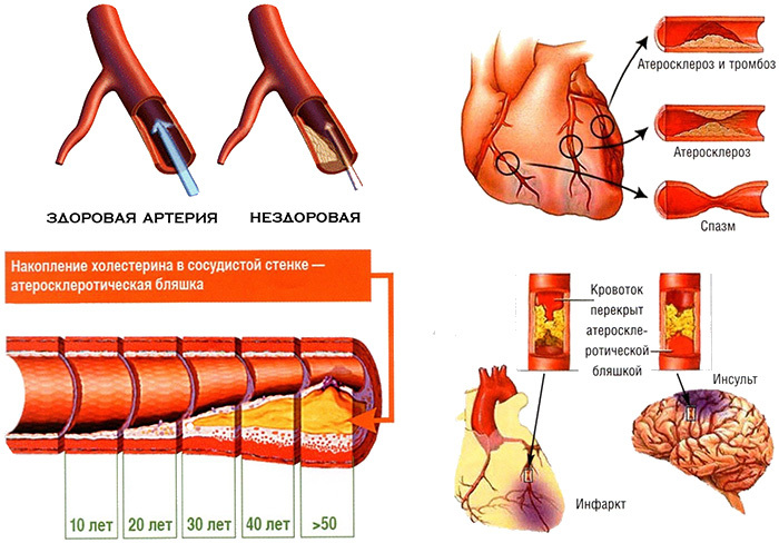
- An artery bulge results from a weakening of the artery walls, which allows blood pressure to "push" them apart. They become wider than usual.
- Aortic aneurysm can form a bulge that is either uniform all the way around the artery ("Fusiform" aneurysm) or protrudes only on one side (saccular or saccular aneurysm).
- Cerebral vascular aneurysm is usually saccular. This form is also characteristic of most cases of ruptured cerebral aneurysms.
- Ruptured cerebral aneurysms are the most common cause of a type of stroke known as subarachnoid hemorrhage. It is less common than ischemic stroke caused by a blockage in an artery rather than internal bleeding.
The mechanism of aneurysm development is not fully understood. Well known only risk factors, which include atherosclerosis. Because of it, 90% of cases of abdominal aortic aneurysm develop. Aneurysms of the ascending thoracic aorta usually develop due to degeneration of the media (middle lining of arterial vessels).
Some aneurysms, although less common, are present as an artery defect at birth (congenital aneurysm). The risk of developing aneurysm and ruptured aneurysm is increased by smoking, excessive alcohol use, and drug abuse, especially cocaine.
Aortic dissection - a tear in the inner wall of an artery - can deform the arterial wall as blood flows between the layers of the aortic walls and stratifies them even more. This condition is called aortic dissection. If the dissection breaks through all three walls of the aorta, it can be fatal. Dissection of the aorta was the cause of death of King George II of England in 1760.
What is a cerebral aneurysm and its symptoms
 A pathology, also known as cerebral aneurysm or intracranial aneurysm, is what a brain aneurysm is. It is characterized by a weak protrusion on the wall of the cerebral artery. The aneurysm in the head is very similar in shape to a small balloon or sac.
As the wall of the artery gradually becomes thinner from dilatation (large accumulation of blood), blood flow causes the weakened wall to swell outward. This pressure can rupture the aneurysm and allow blood to flow into the space around the brain. A ruptured brain aneurysm usually requires surgery.
A small, unexploded (less than a centimeter in diameter) aneurysm in the head is usually completely asymptomatic. Large, unruptured aneurysms can sometimes press on the brain or nerves extending from the brain, causing various neurological symptoms.
These include:
- Frequent localized headache.
- Blurred or double vision.
- Pain above and behind the eyes.
- Difficulty pronouncing words.
A ruptured cerebral aneurysm, as a rule, leads to subarachnoid hemorrhage - bleeding into the subarachnoid space. When blood leaks into the space around the brain, it can cause sudden symptoms.
You should immediately seek medical attention for the following signs of a ruptured aneurysm in the head:
- Sudden severe headache.
- The most severe headache that has ever been in my life.
- Loss of consciousness.
- Nausea and vomiting.
- Torticollis.
- Sudden blurred vision or double vision.
- Sudden pain above or behind the eyes or vision problems.
- Sudden trouble walking or dizziness.
- Sudden weakness and numbness in the limbs.
- Light sensitivity (photophobia) with seizure.
- Hanging eyelid.
Risk factors that contribute to the formation of cerebral aneurysms are smoking, high blood pressure or hypertension, congenital arterial wall abnormalities, family history of cerebral aneurysm, age over 40, gender factors (women compared with men have an increased incidence of aneurysms in the ratio 3:2). Also affected by polycystic kidney disease, Marfan syndrome, the presence of arteriovenous malformations, drug use, aortic infection (vasculitis), tumors or head trauma.
Risk factors that contribute to the rupture of a cerebral aneurysm: smoking, hypertension.
Saccular aneurysm and other types: how is a cerebral aneurysm diagnosed
According to their shape, aneurysms are divided into fusiform (fusimormal) - diffuse expansion of the artery wall over a considerable length and saccular. A saccular aneurysm, as the name suggests, looks like a small sac of blood.
There are 4 types of aneurysm:
- Abdominal aortic aneurysms. These aneurysms are more common than in the past due to the widespread adoption of computed tomography to detect other medical problems. Abdominal aortic aneurysms can be very large and asymptomatic.
- Thoracic aortic aneurysms (AHA). Arises above the diaphragm, a muscle that helps breathe. They do not always cause symptoms, even at large sizes. Because of this type of aneurysm, the valve between the heart and the aorta does not close properly. This allows blood to flow back to the heart. A less common type of AGA can develop in the upper back. Usually occurs as a result of chest trauma, such as from a car accident.
- Brain (cerebral) aneurysms. Also called berry aneurysms because they are often the size of a small berry. Most cerebral aneurysms do not cause any symptoms until they become large or rupture. These are usually saccular aneurysms in shape.
- Peripheral aneurysms. They arise in the arteries and peripheral vessels of the brain extending from the aorta. Common sites for peripheral aneurysms include the popliteal region, thigh region, and carotid artery. Peripheral aneurysms are less prone to rupture or dissection than aortic aneurysms. However, blood clots can also form in peripheral aneurysms. If a blood clot breaks off from the aneurysm, it can block the flow of blood through the artery.
 A brain aneurysm is diagnosed using computed tomography (brain scan). If a CT scan of the skull is performed within 72 hours of the onset of the headache, it can detect 93% to 100% of all aneurysms. In the few cases that are not recognized by CT, the doctor may consider performing a lumbar puncture (SP) to measure blood in the cerebrospinal fluid.
If CT or SP detects the presence of blood, angiography is done to determine where the aneurysm is located. During angiography, a special dye is injected through a catheter into an artery in the brain.
CT is also used to diagnose thoracic and abdominal aneurysms. Also, ultrasound, echocardiography and chest x-ray are used to detect aneurysms.
Important
Aneurysms can be prevented, although they are sometimes congenital. A healthy lifestyle, a normal weight and sleeping at least 7 hours a day can significantly reduce the chance of developing the disease.
What is a ruptured aneurysm and how dangerous it is, what to do to prevent a vessel from bursting in the head
Patients who have an aneurysm and who are aware of the possible complications of this condition often ask doctors the question: "What is a ruptured aneurysm?" This is a rupture of the artery wall with subsequent hemorrhage.
About 10% of patients with ruptured aneurysms die before receiving medical attention. If untreated, another 50% of patients will die within a month, and 25% of patients will have another episode of bleeding within a week. Apart from bleeding issues, there is a significant risk of artery spasm that leads to stroke.
If an aneurysm is found but has not burst, there are two options: either treatment or observation. When deciding which treatment is best for a particular patient, doctors consider several factors. These factors include the patient's age, the size and shape of the aneurysm, the location of the aneurysm, and the patient's neurological condition.
To strengthen the walls of the blood vessels of the brain and prevent the development of aneurysm, as well as the deposition of cholesterol plaques and vascular thrombosis, drugs with trace elements and vitamins are prescribed, such as:
- nicotinic acid - helps to expand capillaries and strengthen the walls of blood vessels;
- vitamin P and ascorbic acid - improve metabolic processes in the walls of arteries and veins, increase their strength;
- silicon, selenium, potassium - trace elements necessary to maintain the tone of cerebral vessels.
 Such drugs are used orally or in the form of injections, courses, after consultation with a neurologist.
Also, a specialist may prescribe antiplatelet agents, fibrates or statins to prevent blood clots, normalize fat metabolism and dissolve atherosclerotic plaques.
After a vessel in the head has burst, either open surgery or an endovascular approach is possible. In a surgical approach, the aneurysm is closed with a metal clip (clip). This prevents blood from entering the aneurysm and subsequent bleeding.
With an endovascular catheter, a catheter is inserted through an artery to the aneurysm and blood flow is blocked in the artery, which ultimately closes the aneurysm.
The main danger of surgical treatment of cerebral aneurysm is the risk of damage to blood vessels and increased bleeding. The result can be spasm of nearby blood vessels and stroke. But sometimes the only alternative to surgical treatment of a burst vessel in the head is death, so doctors and the patient do not have to choose.
note
The prognosis for treatment is generally good. The maximum recovery can take from several weeks to several months.
If the aneurysm was with subarachnoid hemorrhage, the duration of hospitalization and recovery is dictated by the severity of the hemorrhage. After a subarachnoid hemorrhage, most patients remain in the hospital for at least two weeks. Under the best circumstances, many patients can resume their previous activities within a few weeks, without any specific restrictions.
Endovascular therapy, while less invasive, is associated with shorter hospital stays and faster return to previous activities. After a successful endovascular procedure, most patients are seen in the intensive care unit overnight and leave the hospital the next day if there are no complications. These patients can resume their previous activities (including driving) within a few days.
After craniotomy for surgical ligation of the aneurysm, complete recovery takes 4 to 6 weeks. During the recovery period, the patient is able to take care of himself, but does not participate in active physical work.
Can the patient make an independent effort so that the vessel in the head does not burst? Maybe he should.
Important
The most important way to prevent the onset and rupture of an aneurysm is to quit smoking. This habit is a major risk factor for aneurysm-provoking conditions such as atherosclerosis.
Studies have shown that smoking causes a rapid expansion of the aortic aneurysm - by about 0.4 mm per year.
Cigarette smoke is a major source of free radicals that cause oxidative stress and, as a result, tissue damage throughout the body. Oxidative stress can increase inflammation, which contributes to the formation and rupture of cerebral aneurysms.
Almost 80% of patients diagnosed with hemorrhagic stroke due to ruptured cerebral aneurysm were smokers.
It is also necessary to normalize blood pressure - through diet, exercise and, if necessary, medication. It helps to relieve pressure and tension on the walls of the arteries.
Partial use of site materials (no more than 30% of the content of the article) is allowed only with a hyperlink on www.med88.ru, full use of the article (more than 30% of the article content) is possible only with written permission edition.