Ventricular tachycardia: what it is, symptoms and treatment
Content
- What is ventricular tachycardia?
- Consequences
- Epidemiology
- Causes and risk factors
- Symptoms and Signs
- Complications
- Diagnostics
- Treatment
- Prevention and recommendation
What is ventricular tachycardia?
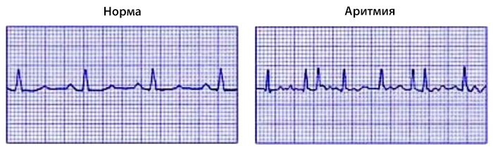
Ventricular tachycardia - cardiac arrhythmiacharacterized by an increase in the heart rate of the ventricles.
The ventricle is a part of the heart, it is divided into the right and left ventricles, which receive blood from the atria and pump it into the arteries.
Stages of development of the disease:
- the ventricles contract too quickly and disorganized in relation to the atria;
- they are not filled in adequately;
- the amount of blood pumped in a circle decreases with each blow;
- the arterial contour decreases;
- the amount of blood that is oxygenated and nourishes the heart also decreases (coronary circulation);
- the contractile efficiency of the heart is further reduced;
- degeneration with ventricular fibrillation;
- death.
This unfortunate evolution is more likely in the case of very high ventricular rates and in the case of cardiac abnormalities in patients with heart failure.
Ventricular tachycardia is one of the most well-known types of arrhythmias in patients with heart disease. Although it can also occur in healthy people, it is an arrhythmia that needs to be treated with caution: in fact, it can develop into ventricular fibrillation, the outcome of which often leads to lethal outcome.
Ventricular tachycardia causes a very strong increase in heart rate. The stomach beats faster, so the number of beats or contractions increases from the normal 60-100 per minute to 150-200 per minute.
Ventricular tachycardia is one of the most common and most dangerous arrhythmias. This is usually a serious heart condition, but it can also occur in healthy people.
Consequences
The regular contraction of the ventricle is responsible for cardiac output. Cardiac output refers to the pumping action of circulating blood on the lungs and tissues of the human body.
The altered ventricular rhythm leads to insufficient cardiac output. Consequently, oxygenated blood does not irrigate the tissues and organs of the body, including the heart, which subsequently loses its contractile efficiency. If this deficiency is severe, the patient faces death.
Epidemiology
The incidence data suggests that:
- Ventricular tachycardia is associated with age: it is more common in middle-aged and elderly people.
- 2-4% of people over 60 years of age without heart problems experience episodes of ventricular tachycardia.
- 4-16% of people over 60 years of age with heart disease experience episodes of ventricular tachycardia.
Read also:Heart diseases
In addition, ventricular tachycardia:
- It is more common during the winter months.
- It has a circadian character: the peak incidence is observed in the morning hours.
Causes and risk factors
The main causes of ventricular tachycardia are heart disease.
This is followed by causes of electrolyte imbalances that alter the electrical activity of the heart.
Finally, there are a number of risk factors that predispose a person to episodes tachycardia.
Most affected by ventricular cardiac tachycardia the patients. Heart diseases seen in these patients:
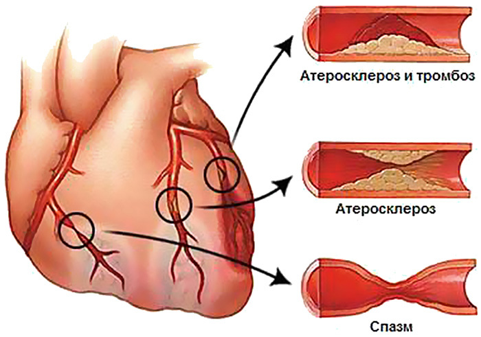
- Coronary heart disease and carried over myocardial infarction.
- Malfunction of one of the heart valves.
- Cardiomyopathy, that is, diseases myocardium (cardiac muscles).
The most common valvulopathies (abnormal heart valve (s)) are those in which the mitral valve is involved.
Cardiomyopathies are rheumatic in nature: in other words, they result from inflammation of the bacterial type. In these cases, we are talking about myocarditis.
A small percentage of cases of ventricular tachycardia are also associated with congenital heart disease. The most famous of them:
- Brugada syndrome.
- Wolff-Parkinson-White syndrome.
Less often, instead:
- Fallot's tetralogy.
- Marfan syndrome.
- Ionic / electrolyte balance.
The pulse of myocardial contraction is an electrical signal. It moves ions that have a positive and negative charge present inside the cells of the heart. The movement of these ions is similar to the movement of charges in an electrical circuit and leads to contraction of the heart muscle.
The main ions with charge are: potassium, magnesium, calcium and sodium. Among them there is an excellent balance, which must be maintained in such a way for the proper functioning of the muscle cell and not only. It may be that this balance has changed. As a result, the contraction pulse decreases and ventricular tachycardia occurs. The main ionic / electrolyte imbalances are:
- Hypokalemia.
- Hypocalcemia.
- Hypomagnesemia.
Risk factors:
- Taking medications.
- Tricyclic antidepressants.
- Cocaine abuse.
- Alcohol abuse.
- Smoking.
- Caffeine.
- Gas poisoning:
- Cyclopropane.
- Carbon monoxide.
- Chest injury.
- Physical and emotional stress.
Symptoms and Signs
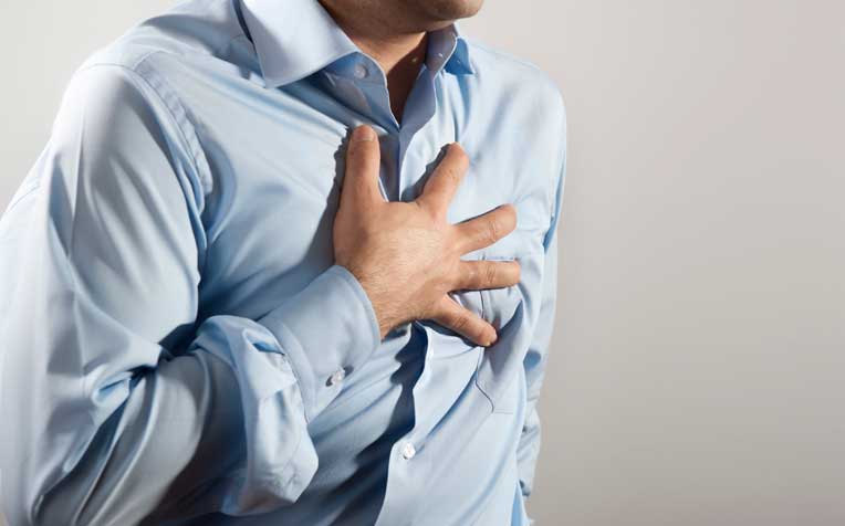
The main symptoms of the disease are as follows:
- Palpitations.
- Chest pain.
- Dizziness.
- Fainting.
- Dyspnea.
In most patients, these symptoms are associated with coronary artery disease or with impaired blood flow (eg, valvulopathy).
The doctor may detect the following clinical signs:
- Accelerated pulse.
- Hypotension.
- Anxiety.
- Excitement.
- Loss of consciousness.
Their appearance depends on the degree of heart damage: the more serious, the easier it is for them to prove themselves.
Complications
Ventricular tachycardia can develop into ventricular fibrillation. This occurs mainly in people with heart disease, while it is very rare in healthy people with paroxysmal ventricular tachycardia.
Read also:Pericarditis, what is it, causes, symptoms and treatment in adults
Ventricular fibrillation is usually fatal. This determines the death of the patient:
- Sudden cardiac death.
- Heart failure.
Diagnostics
Several methods can be used, each of which has a distinct advantage. It:
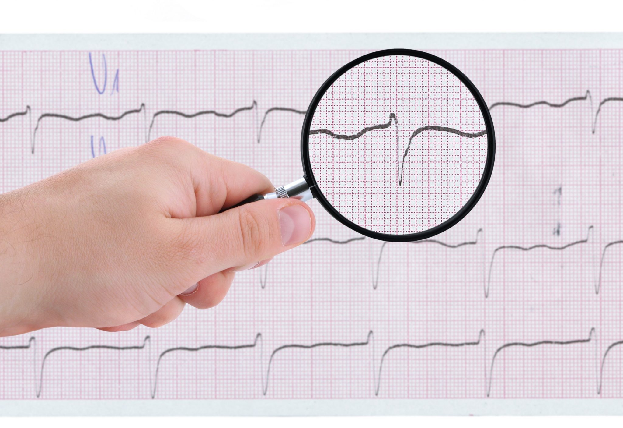
- Electrocardiogram (ECG). It measures the electrical activity of the heart and allows you to identify the form of ventricular tachycardia that affects the patient. It is also possible to monitor cardiac activity for 24 hours; in this case, a dynamic Holter ECG is used. This is a useful study when the form of ventricular tachycardia is paroxysmal, that is, with a sporadic and unpredictable onset.
- Echocardiography. This is a non-invasive test. It uses ultrasound to assess the health of the heart's major structures: the atria, ventricles, and valves. This is useful when valve disease is suspected.
- Chest x-ray. Provides information about the relationship between the heart and lungs. When ventricular tachycardia occurs, pulmonary thrombosis may occur. It is an invasive test because it uses ionizing radiation.
- Coronary angiography. This is an invasive diagnostic method. It is necessary when coronary heart disease occurs at the onset of ventricular tachycardia. Measures the position and degree of occlusion of the coronary arteries in order to plan possible surgical intervention. This is a delicate test as it risks damaging the coronary vessels that are traversed by the catheter.
-
Blood tests. They provide various information about:
- Ion / electrolyte concentration.
- Calcium levels.
- Magnesium levels.
- Phosphate levels.
- The concentration of some drugs taken by the patient.
- Concentrations of some cardiac markers.
Treatment
When heart disease occurs in the onset of ventricular tachycardia, the goal of treatment is twofold:
- Resolve major heart disease. The main goal.
- Arrhythmic disorder solution. Secondary goal.
This is because the second problem is a consequence of the first.
In those who do not have cardiovascular disease, ventricular tachycardia may resolve spontaneously. Thus, the use of the drug can be avoided. In any case, it is advisable to consult a doctor and undergo a thorough examination.
If a patient develops multiple episodes of a persistent type, you can use to block the attack of tachycardia:
- Pharmacological cardioversion.
- Electrical cardioversion.
Pharmacological cardioversion - this is the restoration of a normal heart rate when taking medications:
- Antiarrhythmic drugs to restore normal heart rhythm.
- Lidocaine.
- Amiodarone.
- Procainamide.
- Beta blockers to slow your heart rate.
Electrical cardioversion consists of an electrical discharge to reset and restore normal sinus rhythm.
Read also:Hyperlipidemia
Using a device equipped with two plates applied to the patient's chest. This is also known as defibrillation. Today, there are semi-automatic and automatic defibrillators that can assess the degree of ventricular tachycardia and deliver the correct electrical shock. Another great advantage is that they can be used by non-medical personnel.
Drug therapy is the same as described above. So:
- Antiarrhythmic drugs.
- Beta blockers.
Added to them:
- Anticoagulants to avoid blood clots and embolisms caused by valvulopathies.
In addition to electrical cardioversion, surgery can be performed:
- Transcatheter radiofrequency ablation. A catheter guided into the heart delivers a radio frequency discharge at the point of the ventricle, which generates arrhythmias. The affected area is destroyed, and this should restore a normal heart rate. This is an invasive technique.
- Implantable defibrillator. This is a conventional defibrillator, which, however, is implanted under the skin on the left side of the chest. It is connected to the heart through electrodes that emit an electrical shock when they experience an abnormal increase in heart rate. They work for 7-8 years, after which they need to be replaced. A possible problem could be an instrument malfunction that could cause unplanned electrical discharges.
The therapy to be adopted must, of course, be chosen on a case-by-case basis, not forgetting that the first therapeutic intervention should solve a possible pathological problem that causes ventricular tachycardia.
Prevention and recommendation
Adopting a healthy lifestyle is the best prevention. So, it is recommended:
- Quit smoking.
- Limit alcohol consumption.
- Change your diet.
- Don't use drugs.
- Get exercise.
Tobacco and alcohol are responsible not only for isolated episodes of tachycardia, but also for chronic changes in heart rate. In fact, they are among the most common risk factors for heart disease.
Changing your table habits is another fundamental preventive step. It is advisable to reduce fat, red meat and increase the consumption of fruits and vegetables.
Adopting healthy habits eliminates the possibility of ventricular tachycardia degenerating into ventricular fibrillation. The latter is almost always fatal.
At risk are those who:
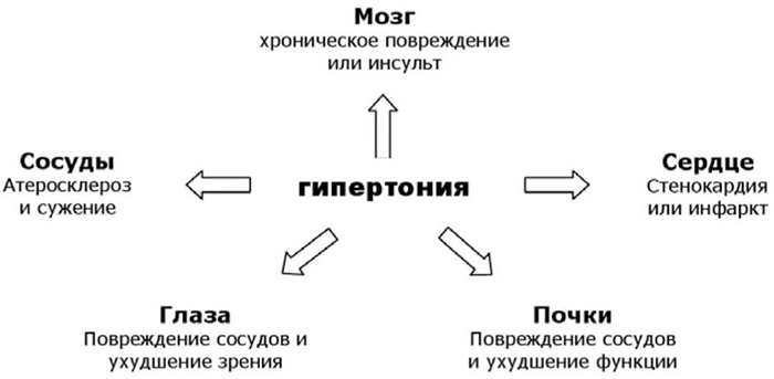
- Represents pathological conditions such as hyperlipidemia, hypertension and diabetes. This contributes to the development of heart disease.
- Has a family history of coronary artery disease.
- Smokes.
- Alcohol abuse.