Macular degeneration: what is it, causes, symptoms, treatment
Content
- What is retinal macular degeneration?
- Types of macular degeneration
- Retinal macular degeneration statistics
- Causes and risk factors for macular degeneration
- Age
- Smoking
- Lack of exercise
- Food
- Heredity
- Genetic factors
- Cataract surgery
- Diabetes
- Possible predisposing factors
- Progression of macular degeneration
- Dry macular degeneration
- Wet (wet) macular degeneration
- Diabetic retinopathy
- Retinal macular degeneration symptoms
- Dry macular degeneration
- Wet macular degeneration
- Diabetic retinopathy
- Clinical examination of macular degeneration
- How is macular degeneration diagnosed?
- Dry macular degeneration
- Wet macular degeneration
- Diabetic retinopathy
- Other eye examinations
- Retinal macular degeneration treatment
- Dry macular degeneration
- Antioxidant Supplements
- Wet macular degeneration
- Vascular endothelial growth factor (anti-VEGF) inhibitors
- Laser therapy
- Macular degeneration surgery
- Radiotherapy
- Diabetic maculopathy
- Macular degeneration prognosis
What is retinal macular degeneration?
Macular degeneration of the retina (macular degeneration
or macular degeneration) Is a disease in which a spot is affected in the central part retinaknown as macula (macula). All over the world, she is the main reason loss of vision and about 50% visual impairment.The condition is characterized by a person's loss of central visual acuity, that is, the part that is used for tasks requiring clear attention, including reading, writing and driving.
As the acuity of central vision deteriorates, a person (what he sees in the center of his field of vision) loses focus, although his peripheral vision (peripheral vision) is seen normally. Although peripheral vision is unaffected, macular degeneration can lead to loss of vision so severe that many people with the condition are considered official. blind.

Types of macular degeneration
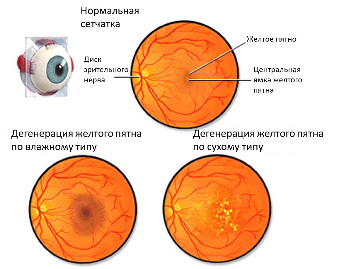
Retinal macular degeneration (macular degeneration) can be classified as:
- Dry macular degeneration (geographic atrophy): Dry macular degeneration is a form of macular degeneration retina that progresses slowly and causes gradual moderate to severe loss vision. Most (90%) cases of macular degeneration are dry;
- Wet macular degeneration (neovascular or exudative macular degeneration): Wet macular degeneration is a form of macular degeneration characterized by a rapid onset. It occurs as a result of a process known as neovascularization (vascular proliferation) that causes ross of abnormal blood vessels under the macular retina. Only about 10% of macular degeneration is of neovascular origin. However, this form of the condition explains much of the blindness associated with macular degeneration;
- Diabetic maculopathy: diabetic retinopathy, or diabetic eye disease, is a condition that affects about a third of diabetic patients. The disease often involves only degeneration of the peripheral retina, which is responsible for peripheral vision. However, sometimes it also affects a person's central vision, causing macular degeneration, in which case it is referred to as diabetic maculopathy.
Retinal macular degeneration also sometimes results from eye trauma, infection, or inflammation.
Macular degeneration (both wet and dry) can also be classified according to the age group of people who develop it.
The vast majority of macular degenerations are observed in the elderly and is partly caused by the aging process known as age-related macular degeneration.
In rare cases, macular degeneration can be of genetic origin and occurs in young people (rarely in children), in medicine this form of the disease is called juvenile macular degeneration or Stargardt disease.
Retinal macular degeneration statistics
Macular degeneration usually affects older people over 50 years oldin whom visual acuity (visual acuity) decreases slowly in a natural way. Globally, this is the main root cause of vision loss (blindness) and about 50% of visual impairment.
In Russia, this is the main cause of serious visual impairment in the elderly. About 15% of the population at the age of 50, symptoms of early macular degeneration appear. In the early stages, the condition does not affect a person's vision, but there is an increased risk of developing late or progressive macular degeneration of the retina, resulting in severe vision loss.
The risk of vision-threatening macular degeneration increases exponentially with age. In the age group 40-49 years old the prevalence is 0.8%, compared with 16% over the age of 90.
For 20 years, diabetic retinopathy affects an estimated one third of the world's people with diabetes. About 7.5% of people have diabetes, so in twenty years, 2.5% of the population will suffer from diabetic retinopathy. However, it is unclear what proportion of these cases are associated with macular retinal degeneration, and what proportion only with peripheral retinal degeneration.
In rare cases, macular degeneration can develop in young adults, and almost always due to genetic predisposition.
Causes and risk factors for macular degeneration
Age
The risk of retinal macular degeneration increases as a person gets older. The disease usually affects people in their 50s.is called age-related macular degeneration.
The risk increases exponentially with age. Old people aged 90 have a 25% chance of severe vision loss due to macular degeneration versus 1-2% risk elderly people aged 50 years.
Smoking
Smoking is a well-established modifiable risk factor for macular degeneration. For example, results from three large cross-sectional studies have shown that smokers are more than 3 times more likely to develop age-related macular degeneration than nonsmokers.
There is also evidence that the more a person smokes, the higher the risk of macular degeneration.
Lack of exercise
Available evidence suggests that regular physical exercise reduce the risk of macular degeneration in humans, and therefore lack of exercise increases the risk of macular degeneration.
For example, one study found that people who exercised for at least 30 minutes three times a week were more likely to have macular degeneration decreased by 70%than those who did not. However, it is unclear whether this link is direct or indirect, as a result of other health benefits of regular exercise (such as improved cardiovascular health).
Food
There is evidence that a number of factors affecting nutrition affect the progression of age-related macular degeneration (how quickly vision deteriorates in a person with such disease).
For example, one study reported that individuals without diabetes but with a high glycemic index (a measure of the proportion of carbohydrates in the diet and the degree to which which they raise blood sugar) are more likely to develop age-related macular degeneration than those with a low glycemic index.
Read also:Aniridia
Eat a healthy, balanced diet. Limit your fat intake, eat fatty fish two to three times a week, eat dark green leafy vegetables and fresh fruits daily, and a few nuts a week.
Heredity
People with a family history of macular degeneration are at a higher risk of developing the condition than those without a family history of macular degeneration. One study reported more than 10 times an increased risk of macular degeneration in individuals with siblings with the condition. Another study found that there is a 50% chance of developing macular degeneration if there is a family history of macular degeneration.
Genetic factors
Some people are genetically predisposed to retinal macular degeneration, and they usually have the disease develops at an early age, sometimes during childhood. Macular degeneration of genetic origin is not well understood, but it is believed that it is associated with the mutation of a number of genes. However, further research is needed to determine the effect of the gene mutation on the development of genetic macular degeneration.
Genetic diseases associated with early macular degeneration include:
- Best's disease (a condition characterized by an accumulation of yellow pigment around the macula that causes cell damage);
- Stargardt's disease (a genetically inherited form of macular degeneration that affects children aged 7-12);
- Sorsby dystrophy (a genetic disorder affecting children with characteristics similar to wet macular degeneration).
Cataract surgery
Two large foreign studies have shown an increased risk of macular degeneration in people who have had surgery to remove it. cataracts, compared to those who did not.
First, Australian study reported a more than 3-fold increased risk of wet and dry macular degeneration in patients who underwent cataract surgery compared to patients who did not.
Other study in the United States also reported a more than 3-fold increased risk of late vision-threatening macular degeneration in patients undergoing cataract surgery.
Diabetes
Diabetes is a key risk factor for diabetic retinopathy. One third of patients diabetes mellitus retinopathy develops 20 years after the onset of diabetes, and some of these cases are associated with macular degeneration (others with peripheral retina, but not macular).
Patients with diabetes mellitus are at greatest risk, arterial hypertension and hyperlipidemia (abnormally elevated blood lipids) that are poorly controlled.
Possible predisposing factors
Research has shown that the following factors may have an association between a specific risk factor and the risk of developing retinal macular degeneration:
- Hypertension: Several studies have found a link between hypertension and macular degeneration.
- Factor H: There is new research that there is a link between complement factor H and the development of macular degeneration. Complement factor H is an important part of the complex protein system, which in turn is an integral part of the body's immune system.
- Blue eye color.Blue color of the iris is associated with age-related macular degeneration.
- Exposure to sunlight: A link has been found between exposure to sunlight and the development of macular degeneration. This link has been attributed to high levels of exposure to sunlight, causing damage to the eyes, which can then lead to macular degeneration of the retina at a later age.
Progression of macular degeneration
The onset and progression of macular degeneration varies by type.
Dry macular degeneration
Dry macular degeneration is characterized by slow onset and progression. The condition usually remains asymptomatic for many years and can affect one or both eyes.
Dry macular degeneration due to geographic atrophy (destruction of blood vessels) in the retinal area and destruction of the retinal epithelial pigment, the membrane that separates the choroid and the retina.
Patients with damage to only one eye may compensate for this with their good eye, and not realize that their vision has been deteriorating for a longer period of time. In cases of dry macular degeneration, vision loss progresses slowly and often develops over the years.
Wet (wet) macular degeneration
Wet macular degeneration is a rapidly progressive condition. Approximately in 30% of people with wet macular degeneration, only one eye is affected, but there is a high risk (40% over 5 years) of affecting the other eye.
If untreated, wet macular degeneration results in rapid visual impairment; patients lose an average of three lines of visual acuity (measured by checking which lines of smaller letters a person can see on an eye map) after a year.
Wet macular degeneration occurs due to choroidal neovascularization, or abnormal growth of neovascular membranes (microscopic blood vessels) beneath the macular gland. She causes bleeding and scarring of the retina and irreversible damage to photoreceptors (which receive and process light rays entering the eye).
Choroidal neovascular the membrane arises in the choroid (the part of the eye located between the retina and the sclera) and breaks through the retinal pigment epithelium, which separates the choroid and the retina.
Diabetic retinopathy
Diabetes-related retinopathy results from chronic hyperglycemia (elevated blood sugar), which causes a number of changes in the blood vessels. These include thickening of the retinal membrane and neovascularization in the macular region, similar to that which occurs with wet macular degeneration.
These changes result in decreased blood flow in the capillaries (small blood vessels) that feed the retina (including macular), and bleeding in the vitreous humor, a gel-like substance that fills the area of the eye between the lens and retina. These changes cause vision loss and ultimately lead to complete blindness if left untreated.
Retinal macular degeneration symptoms
The main symptom of retinal macular degeneration is the loss of visual acuity in the central part.

The exact visual changes that people experience as they develop retinal macular degeneration vary with the type of macular degeneration.
If you suspect you have macular degeneration, it is important to write down the exact nature of the visual changes, including when they started, how often they occur, and how vision is impaired. The doctor will be interested in these changes if he suspects macular degeneration, additional details will assist the doctor in making a diagnosis.
Dry macular degeneration
Dry macular degeneration is characterized by the following symptoms:
- Gradual loss of visual acuity: In the early stages of the disease, it is usually difficult to read the finely written letters in newspapers or the need for brighter reading lights. In more advanced stages, people with dry macular degeneration may experience blurred vision and difficulty judging distances.
- Distorted color sensitivity: colors appear dull or intelligible, or there may be less contrast between them.
Read also:Rupture of eye capillaries
People who experience these symptoms should visit a general practitioner to assess their eye health. If necessary, the therapist will be able to refer you to an ophthalmologist (optometrist).
Wet macular degeneration
Wet macular degeneration is characterized by the following symptoms:
- Rapid loss of visual acuity: often leading to distorted vision, for example, a person may begin to see straight or wavy lines and have difficulty reading small print;
- Central scotoma: Visually blind areas in the visual field that become larger if left untreated.
Individuals experiencing the above symptoms should see a doctor immediately. Signs point to wet macular degeneration, which leads to rapid vision loss. Diagnosis of the disease in the early stages is vital, since after loss of vision, it cannot be restored.
Treatment for this form of the condition is only effective in preventing further vision loss. The general practitioner will be able to assess the symptoms and if wet macular degeneration is suspected, the person will be referred for an urgent examination by an ophthalmologist.
Diabetic retinopathy
Diabetic retinopathy is characterized by the following symptoms:
- decreased central visual acuity;
- loss of color sensitivity, especially the ability to see yellow and blue;
- light flashes and flickering before the eyes.
Clinical examination of macular degeneration
If there is a suspicion of retinal macular degeneration, a doctor or ophthalmologist will check the person's visual acuity using eye tests. Surveys that can be carried out:
- Standard pungency test view on the table (diagram) with the letters: the method of examination of visual acuity, in which people close one eye and read from the Sivtsev table, containing letters of different sizes (W, B, M, H, K, S, I);
- Amsler grid (Amsler grid): The Amsler grid is a grid of intersecting vertical and horizontal lines with a dot in the middle. People look at a dot and indicate any lines that appear blurry or faded. Vision of blurry or faded lines indicates macular degeneration;
- Color vision test: color sense test is an examination that assesses a person's ability to distinguish between different colors.
How is macular degeneration diagnosed?
To diagnose retinal macular degeneration, your doctor will examine your eyes using a microscope. While the doctor is doing the exam, a very bright light, called a slit lamp, will be directed into the eye. Different types of macular degeneration cause different microscopic changes in the eye, and the doctor will look for them when examining the eye.
Dry macular degeneration
When the following signs are found on examination, it indicates that the person is developing dry macular degeneration:
- hyper- or hypopigmentation of the retina (areas of the retina are noticeably darker or lighter than the rest);
- macular discoloration of the macula;
- large fat deposits around the macula;
- soft body fat;
- destruction of the retinal pigment epithelium (the membrane that separates the retina from the choroid and supplies it with nutrients).
Wet macular degeneration
When the following signs are found on examination, it indicates that the person is developing wet macular degeneration:
- subretinal fluid (accumulation of fluid around the retina);
- subretinal hemorrhage (hemorrhage around the retina);
- scarring of the retina.
Diabetic retinopathy
When the following signs are found on examination, it indicates that the person is developing diabetic retinopathy:
- microaneurysms (stretching of microscopic blood vessels);
- hemorrhage;
- Hard exudates (small, yellow deposits that form from blood vessels flowing into the retina)
- change in veins;
- the formation of new veins;
- thickening of the retina.
Other eye examinations
If the examination reveals retinal macular degeneration, other examinations may be performed:
- Fluorescence angiography: X-ray of the eye, carried out after the introduction of a fluorescent dye into the blood vessels. This test can be used if wet macular degeneration is suspected. This allows the doctor to assess the extent, size, and location of neovascularization (abnormal growth of blood vessels) and the extent to which blood flows out from the blood vessels. An examination can also be performed to exclude non-macular degenerative causes of visual impairment;
- Optical coherence tomography: a technique that uses a laser to create an image of the eye or other organs. It is used to assess the degree of retinal thickening and fluid accumulation in the retinal tissue.
- Indocyanine green angiography: a procedure similar to fluorescence angiography, but using a different type of dye, also can be used to help a doctor assess the extent of abnormal growth of blood vessels. This test is usually not necessary as fluorescein angiography will provide this information.
Retinal macular degeneration treatment
Treatment for macular degeneration is prescribed to prevent further loss of vision. Treatment varies depending on the type of macular degeneration.
Dry macular degeneration

There are currently no treatments available to prevent further vision loss from dry macular degeneration.
Therapy is aimed at controlling risk factors for the disease. To reduce further vision loss from dry macular degeneration, people should:
- quit smoking;
- control blood pressure;
- eat foods rich in fruits and vegetables;
- wear a hat and sunglasses when exposed to sunlight;
- lead a healthy lifestyle, control your weight and exercise regularly.
However, it should be noted that there is no scientific evidence that control of blood pressure or exposure to sunlight causes progression of macular degeneration. These are precautions.
People should also take steps to reduce the likelihood of vision loss that progressively worsens:
- undergo regular examinations;
- improve lighting in the house;
- use magnifiers and reading lamps;
- use other visual enhancers.
Antioxidant Supplements

It has been suggested that taking antioxidant supplements protects a person from retinal macular degeneration, although scientific evidence that antioxidant supplements prevent or delay the onset of disease no.
However, there is evidence that high doses of antioxidant and zinc supplements reduce further vision loss in cases of progressive macular degeneration.
Wet macular degeneration
Controlling risk factors and adapting to vision loss are equally important components of wet macular degeneration therapy. There are also a number of treatments available to prevent and reverse neovascularization. They are effective in preventing further loss of vision, and in some cases, partially restore vision.
Vascular endothelial growth factor (anti-VEGF) inhibitors
Vascular endothelial growth factor inhibitors target a substance in the body called vascular endothelial growth factor (VEGF). VEGF is the main stimulant of choroidal neovascularization or abnormal growth of blood vessels in wet macular degeneration, therefore blocking the action of VEGF prevents neovascularization.
Read also:Double vision (diplopia): causes and treatment
Vascular endothelial growth factor (anti-VEGF) inhibitors Are drugs that effectively prevent and sometimes improve vision loss in patients with wet macular degeneration.
Ranibizumab (Lucentis)
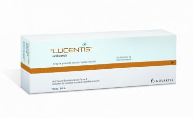
Lucentis is a drug that targets and binds to VEGF type A. In doing so, it prevents VEGF-A from binding to its receptors VEGF-1 and VEGF-2, which in turn prevents abnormal growth of blood vessels.
Scientific research has shown that Lucentis is very effective in preventing and improving vision loss in a significant proportion of patients who have received therapy.
In two studies About 95% of patients who received Lucentis injections retained their vision for two years, compared with 62% of people who did not receive the drug. About a third of those who took Lucentis experienced an improvement in visual acuity compared to 5% of those who did not take the medication.
Laser therapy
There are two types of laser therapy that are used to treat retinal macular degeneration, and about a third of people with this condition can benefit from laser therapy.
Photodynamic laser therapy is the preferred method because it causes less damage to the retina than conventional argon laser therapyThis is another type of laser treatment for macular degeneration.
Photodynamic laser therapy
Photodynamic laser therapy is the introduction by a physician to a sick patient of a photosensitizing dye, that is, a dye that is sensitive to light rays.

The part of the eye damaged by abnormal growth of blood vessels will absorb most of the this dye, which will eliminate the effect of the dye on the blood vessels in the surrounding retina.
The dye is activated by a low power diode laser that targets the eye. When the rays of light enter the eye, they activate a photosensitizing dye, which will then only destroy cells that have absorbed the dye (cells of damaged blood vessels that cause yellow degeneration spots).
Other cells in the retina will not be destroyed because they will not absorb the dye. This is a pretty smart and effective therapy.
People will need to undergo regular examinations (three times a month) after treatment, therapy is usually requires 3-5 sessionsto completely eliminate the abnormal growth of blood vessels.
Macular degeneration surgery
Macular degeneration surgery consists in detaching the retina and moving it to the part of the eye that is not damaged by abnormal growth of blood vessels. There is currently limited evidence for the effectiveness of this procedure.
Available evidence suggests that it carries a high risk of side effects. For example, more than 20% of people who undergo surgery have retinal detachment. Therefore, retinal macular degeneration surgery is not recommended as a routine treatment for wet macular degeneration.
Radiotherapy
There is some evidence that external bean radiation therapy is effective in preventing abnormal growth of blood vessels in the retina. However, because this treatment has many serious side effects, it is not recommended.
Diabetic maculopathy
The following treatments are effective in preventing macular degeneration in diabetes-related cases:
- Focal laser coagulation (photocoagulation) of the retina. The procedure, in which the blood vessels are affected by a powerful laser, the procedure lasts 20-30 minutes, is an effective treatment in cases of proliferative degeneration accompanied by severe macular swelling. This treatment is more effective in treating diabetic eye diseases affecting the peripheral retina;
- Triamcinolone acetonide (Kenacort A10): the drug Kenacort is injected into the eye, similar to Lucentis. It improves visual acuity and decreases macular thickness in patients with diabetic macular edema (excessive fluid accumulation). However, it should be noted that this treatment is off-label, that is, the drug is not approved for the treatment of eye diseases. Treatment usually includes repeated injections and significant adverse reactions.
Macular degeneration prognosis
Retinal macular degeneration is characterized by progressive loss of vision, which is slow in cases of dry macular degeneration and rapid in cases of wet macular degeneration.
Treatment options depend on the type of macular degeneration. Usually, vision cannot be restored and treatment is aimed at preventing further deterioration in vision.
There is currently no effective treatment for dry macular degeneration. In these people, early detection and prevention of further vision loss is a key strategy for treating this condition.
There are several treatments that are effective in preventing vision loss and in some cases for partial restoration of vision in persons with wet macular degeneration (outlined in outline higher). Due to the rapid onset of the condition, treatment should be started quicklybefore scarring appears on the retina. Therefore, early detection is vital.
There are also treatments that can prevent vision loss in cases of diabetic maculopathy.
Retinal macular degeneration is a painless condition, but it leads to loss of vision. Thus, the condition has significant implications for the quality of human life. Although macular degeneration does not cause complete blindness, because people retain their peripheral vision, vision loss is common. so severe that individuals with progressive macular degeneration are officially classified as blind and given disability.
Loss of vision can prevent a person from performing previously accepted taskssuch as driving, reading and personal grooming. They may need to change aspects of their lifestyle, for example, they may need to walk or ride public transport instead of driving, or become addicted to someone else's care.
People may be anxious about the threat of total blindness, and where treatment is possible, it is unpleasant. (for example, with injections into the eyes), requires frequent clinic visits and often leads to side effects therapy.
A significant proportion of people with macular degeneration (30%) experience emotional distress, including depression and anxiety resulting from loss of independence and difficulty in performing basic tasks that result from loss of vision.
Approximately 12% of people experience a syndrome known as Charles Bonnet syndrome, a visual disorder characterized by visual hallucinations. They are often reluctant to discuss their hallucinations with a healthcare professional for fear of being perceived as mentally ill. Loss of vision is also associated with a higher incidence of falls and, with it, morbidity (eg, fractures).