Amniotic fluid embolism: what is it, causes, symptoms, treatment
Content
- What is amniotic fluid embolism?
- Signs and symptoms
- Causes
- Affected populations
- Similar disorders
- Diagnostics
- Clinical testing and examination
- Standard treatments
- Forecast
- Prospects for moms
- Prospects for children
What is amniotic fluid embolism?
Amniotic fluid embolism (EOV) or anaphylactoid pregnancy syndrome (ASB) is an extremely rare but life-threatening complication that affects pregnant women shortly before, during, or immediately after childbirth. Most cases occur during childbirth.
It is assumed that with this disease, the pregnant woman has a severe allergic reaction to the amniotic fluid (amniotic fluid, fetal fluid) or other fetal materials such as embryonic cells that enter mother's blood flow. Amniotic fluid is contained in the amniotic sac. This liquid supports, softens and protects the developing fetus. Amniotic fluid embolism is unpredictable and no risk factors have been identified. Anaphylactoid pregnancy syndrome can cause serious, rapid deterioration in the health of the mother.
For years, some researchers believed that amniotic fluid and fetal cells caused obstruction in the mother's blood vessels, but now most researchers believe that the mother's immune system reacts to the amniotic fluid and fetal cells, causing an overwhelming immune system response that ultimately harms the mother and the fetus.
Breathing problems, cardiac arrest, and excessive bleeding are some of the life-threatening complications that can occur. Researchers and doctors do not fully understand why amniotic fluid or fetal cells that enter the mother's bloodstream cause this reaction in some women. Amniotic fluid embolism is a medical emergency that develops suddenly and quickly and can be fatal. Early detection and prompt supportive care are essential.
Signs and symptoms
Signs and symptoms of amniotic fluid embolism often develop rapidly. Nonspecific symptoms including headache, chest pain, coughing, sweating, nausea and vomiting were reported as early signs.
Other common initial symptoms include:
- labored breathing (dyspnea);
- abnormally fast breathing (tachypnea);
- low blood pressure (hypotension);
- abnormally fast heartbeat (tachycardia);
- low oxygen content in the body (hypoxia);
- cyanotic discoloration of the skin and mucous membranes due to lack of oxygen (cyanosis) in blood;
- a rapid increase in blood pressure in the blood vessels of the lungs (pulmonary hypertension);
- sudden narrowing of blood vessels (vasospasm).
Breathing problems can develop into acute respiratory failure - a serious, life-threatening complication where damage and leakage fluid in the lungs make breathing difficult or impossible. Some affected women experience increased heart rate, low blood pressure, shock and / or a sudden loss of heat function despite the absence of underlying heart disease (cardiac arrest hearts). Cardiac arrest can cause labored breathing, lack of breathing or complete immunity of the affected person. Affected women may experience mental status changes, including anxiety or confusion, seizures, or coma.
Most women condition developscalled disseminated intravascular coagulation (DIC syndrome). In DIC, blood clotting factors, specialized proteins that help blood clot, are destroyed. Without these clotting factors, sick women do not form blood clots that stop bleeding. There may be bleeding from the uterus and from puncture sites such as the site of an intravenous needle or epidural catheter. In rare cases, heavy bleeding may be the first sign of amniotic fluid embolism..
Doctors with PSP can identify the condition, which is called a pronounced violation of the condition of the fetus. This term means that the fetus has problems. The fetus may not be getting enough oxygen and an abnormally low heart rate may be observed. The Fetal Health Index (FFI) is an analysis of the condition of the fetus at the end of pregnancy or during labor. In the event that anaphylactoid pregnancy syndrome occurs during pregnancy, childbirth should be taken urgently as a saving measure for both the mother and the fetus.
Read also:Down syndrome: what does the extra chromosome carry?
Causes
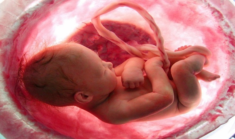
Amniotic fluid embolism is a condition that occurs due to the fact that there is a systemic reaction, similar to the one which is found in a pregnant mother with an allergic response to amniotic fluid or fetal cells or foreign bodies fetus. Amniotic fluid and other materials enter the mother's bloodstream, most likely due to small tears in the lower part of the uterus, the part of the cervix that forms the canal that connects the vagina to the uterus (endocervix), or due to injury or abnormality placenta.
The cervix is the narrow passage that forms the lower end of the uterus. The placenta is the organ that connects the developing fetus to the mother. The placenta allows oxygen and nutrients to be transported from the mother to the fetus.
How amniotic fluid or fetal cells cause an allergic reaction in some women and not others is not entirely clear.
Initially, researchers believed that amniotic fluid had an embolic effect, which means that amniotic fluid forms clots (embolisms) in the mother's blood vessels, most likely in the blood vessels lungs. Hence the name of the disorder - amniotic fluid embolism. While amniotic fluid or fetal cells may contribute to some mechanical obstruction, amniotic fluid dissolves in fluid, and fetal cells or tissue debris that can also enter the mother's blood are usually too small to form clot.
Researchers now believe that it is an immune-mediated disorder and that the symptoms are caused by immune maternal system that reacts to amniotic fluid or fetal cells as foreign substances. This immune response triggers an overwhelming inflammatory response in the mother's body, damaging healthy tissues and structures, ultimately causing the signs and symptoms of the condition. The process is more like anaphylactic shockthan embolism.
While many potential risk factors have been cited, there is strong evidence that any of the risk factors increase the risk of anaphylactoid pregnancy syndrome in there are no women. Most of the studies were inconsistent or conflicting regarding risk factors.
Doctors can neither predict nor anticipate the occurrence of amniotic fluid embolism, and even more so, prevent this disorder.
Affected populations
Amniotic fluid embolism is a rare complication that occurs just before, during, or immediately after birth. The number of women who have experienced this complication is unknown.
Amniotic fluid embolism is a rare condition. Rare disorders often go unrecognized or misdiagnosed, making it difficult to determine their true frequency in the general population. Estimates ranged from 1 in 8,000 to 1 in 80,000 pregnancies.
Similar disorders
Symptoms of the following disorders may be similar to those of amniotic fluid embolism. Comparisons can be useful for differential diagnosis.
There are many different conditions or disorders that can cause symptoms or signs similar to those seen with amniotic fluid embolism. These include:
- cramps during pregnancy due to high blood pressure (eclampsia);
- separation of the placenta from the wall of the uterus (placental abruption);
- heart failure during the last few months before or after birth (peripartum cardiomyopathy);
- anaphylaxis;
- air embolism;
- The entry of material from the stomach into the lungs or lower respiratory tract (pulmonary aspiration);
- shock due to extremely rapid low blood pressure caused by widespread infection (septic shock);
- loss of more than 20% of the blood supply (hemorrhagic shock), which can be caused by several events, including a ruptured or kinked uterus;
- heart attack (myocardial infarction).
Read also:Vesicoureteral reflux (VUR) in children
Diagnostics
The diagnosis of amniotic fluid embolism is based on identifying only characteristic clinical symptoms. To date, there are no diagnostic tests, imaging studies, or pathological markers that can confirm the diagnosis of EOS.
Prompt diagnosis and early aggressive advanced life support methods are essential to ensure the best possible outcome for amniotic fluid embolism. There is no internal consensus in the medical community regarding diagnostic criteria for this disorder, although diagnostic criteria have been proposed for reporting EOV studies (Clark SL, 2016).
Diagnosis is made by identifying characteristic symptoms and excluding other possible causes of signs and symptoms (differential diagnosis).
Clinical testing and examination

Various tests can be ordered that can help treat amniotic fluid embolism. These tests include complete blood countwhich can indicate low hemoglobin levels or high levels of certain heart enzymes. Can be carried out blood coagulation testswhich will show low platelet and fibrinogen levels, increased prothrombin time and activated partial thromboplastin time (APTT).
Platelets Are specialized red blood cells that help blood to clot. Fibrinogen Is a special protein found in the blood. One of the clotting factors that is needed to help blood clot. Prothrombin time (PTT) and APTT Are tests that determine how long it takes for blood to clot.
Two different tests can be used to determine low oxygen levels - continuous pulse oximetry and arterial blood gas analysis. Continuous pulse oximetry is a simple test in which a small sensor is attached to a fingertip or earlobe to measure the amount of oxygen in a person's blood. Your doctor may take a small blood sample for an arterial blood gas test, which can evaluate both oxygen and carbon dioxide levels in your blood.
Chest x-ray can show fluid in the lungs, a noticeable pulmonary artery, or an enlarged heart (cardiomegaly). Electrocardiogram, echo or echocardiographyAre tests that use high-frequency sound waves to create images of the heart. Tests can show heart function, abnormalities and irregularities in the heart rhythm, and dysfunction of the right or left upper chamber of the heart (ventricular dysfunction).
Sometimes doctors took a pulmonary blood sample to look for fetal squamous cells that were believed to be covered in white blood cells called neutrophils and fetal debris such as tissue or cells fetus. Once this analysis was performed to diagnose amniotic fluid embolism, but it is no longer considered compelling evidence for this disorder, since these data are not specific to this disorders.
Standard treatments
Amniotic fluid embolism is a medical emergency. Treatment is aggressive and supportive. A team of specialists is needed to treat the affected individuals. This group will include:
- pain management and patient care specialists before, during and after surgery (anesthesiologists);
- doctors specializing in pregnancy, childbirth and the female reproductive system (obstetricians);
- obstetricians specializing in maternal and fetal health (maternal and fetal medicine specialists);
- doctors who specialize in the diagnosis and treatment of blood diseases (hematologists);
- physicians specializing in the treatment of critically ill patients (intensive care physicians).
Read also:Shereshevsky-Turner syndrome - causes, symptoms, diagnosis and treatment
Specific therapeutic procedures and interventions can vary depending on numerous factors, such as:
- the severity of the disorder;
- the presence or absence of certain symptoms;
- whether the patient is conscious, breathing, etc.;
- the person's age and general health;
- health and condition of the fetus;
- and other factors.
Initially, doctors will work to stabilize the woman's breathing and heart function. If the affected woman is unconscious, she may receive cardiopulmonary resuscitation (CPR). The affected woman may receive additional oxygen to make up for the lack of oxygen in the blood or tissues. This may require the placement of a breathing tube to facilitate breathing and the use of a breathing apparatus (mechanical ventilation).
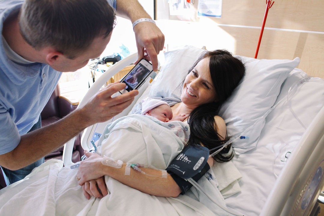
Sometimes doctors insert a small, thin tube called a catheter into the main artery of the lungs (pulmonary artery catheter). This catheter can be used to monitor blood pressure. A central venous catheter can be placed in a large vein and used to deliver medication, draw blood, transfuse blood, or deliver fluids or nutrients.
Some women may get medicine to improve heart function or reduce fluid buildup in or around the lungs. Medicines that cause the blood vessels to narrow are called vasopressorscan be used to treat low blood pressure (hypotension) and improve blood pumping (cardiac output). Medicines that change the speed or strength of muscle contractions, called inotropic drugscan also be given to improve cardiac output. Medications may also be prescribed to maintain the muscle tone of the uterus. Sometimes there is a loss of muscle tone called uterine atony.
Some women will need blood transfusionto replace lost blood and replace clotting factors, which are necessary for blood to clot. Most patients require activation of hospital-specific bulk transfusion protocols, since women who have had anaphylactoid pregnancy syndrome require a multicomponent therapy.
Early labor is essential for the health of the fetus. Reducing aorto-caval compression may improve maternal resuscitation.
Psychosocial support for the entire family is also important. Some women experience amniotic fluid embolism without any long-term complications of the condition. However, there is a risk of long-term complications, including neurological problems, due to lack of oxygen in the brain.
Forecast
Prospects for moms
Estimated mortality rates for women with anaphylactoid syndrome, pregnancy varies greatly. Earlier reports indicate that up to 80 percent of women do not survive, although the most recent figure is around 40 percent. Women who survive this condition often have long-term complications. They can include:
- memory loss;
- organ failure;
- damage to the heart, which can be short-term or permanent;
- problems with the nervous system;
- partial or complete hysterectomy;
- damage to the pituitary gland.
Emotional problems can also arise, especially if the child does not survive. These problems include postpartum depression and post-traumatic stress disorder.
Prospects for children
Estimates of mortality rates for children with EOB also vary greatly. 30 percent of babies do not survive amniotic fluid embolism, reported Journal of Anesthesiology Clinical Pharmacology). However, the EOV Foundation reports that the mortality rate for babies still in the womb is as high as 65 percent. Some babies who survive have long-term or lifelong complications after birth. They include:
- a disorder of the nervous system, which can be mild or severe;
- lack of oxygen in the brain;
- cerebral palsy, which is a disorder that affects the brain and nervous system.