Pneumothorax: causes, symptoms and treatment
Pneumothorax is a disease that causes air to accumulate in the pleural space of the lungs. Their depressurization occurs.
Air penetration into the pleura increases pressure. Then there is a partial or complete recession of the lung.
The human condition is very grave and needs urgent help. Pneumothorax is open and closed. Its occurrence is often due to lung diseases or injuries received (stab wounds, bullet wounds, and so on).
Content
- Causes of the disease
- Symptoms
- Diagnostics
- Complications
- Treatment
- Medication (medications)
- Surgery
- Postoperative exercise
- Complementary and alternative home treatments
- Herbal treatment
- Prophylaxis
- Forecast
- Related Videos
Causes of the disease
The likelihood of developing spontaneous pneumothorax is observed in middle-aged people. The reasons for the occurrence can be:
- bullous disease;
- infectious diseases (atypical pneumonia, pulmonary tuberculosis);
- endometriosis of the lungs;
- interstitial lung lesions;
- malignant formations;
- inflammation of the connective tissue (ankylosing spondylitis and Marfan's disease, rheumatoid arthritis, polymyositis).
Traumatic pneumothorax is formed after trauma to the chest cavity. Distinguish:
- Penetrating chest wounds (stab wounds, gunshot wounds, as well as shrapnel).
- Breast trauma without a penetrating effect from the external environment (provoked by tissue trauma by the sharp edges of broken ribs, lung rupture).
Valvular pneumothorax forms after spontaneous or traumatic. It is one of the most dangerous species.
Iatrogenic pneumothorax can be a consequence of medical procedures. Such as:
- puncture of the pleura;
- improper placement of a central vein catheter;
- taking a sample of the affected lung tissue (biopsy);
- Endoscopic transbronchial biopsy;
- rupture of the alveoli during mechanical ventilation of the lung (barotrauma).
Artificial pneumothorax is used to treat tuberculosis (mainly with fresh destructive forms). This is a procedure for introducing oxygen into the pleural cavity. The procedure is used in order to reduce the formed cavities.
Pneumothorax in newborns is considered abnormal. The occurrence of this disease is associated with genetic pathologies of the lungs and pleura, as well as trauma and inflammatory processes. The reasons may be:
- crying heavily;
- rupture with forced artificial respiration;
- genetic pathology;
- ruptured lung abscess;
- rupture of the cyst.
Catamenial or menstrual pneumothorax is a rare form that develops 2-3 days after the onset of menstrual bleeding. Occurs for reasons:
- intrathoracic endometriosis;
- production of a hormone during ovulation - prostaglandin F2. Its appearance causes narrowing of the bronchioles.
- the absence of a mucous plug in the cervix, which allows air to pass through the openings of the diaphragm into the pleura.
Symptoms

The symptoms of pneumothorax appear due to the accumulation of air in the pleural cavity. Their development depends on the stages of lung reduction.
The size of the collapse of the lung is divided into:
- small (up to 25%);
- medium (50-70%);
- total (100%);
- tense (displaced mediastinum).
Spontaneous pneumothorax is:
- primary (idiopathic);
- secondary (symptomatic);
- recurrent appearance.
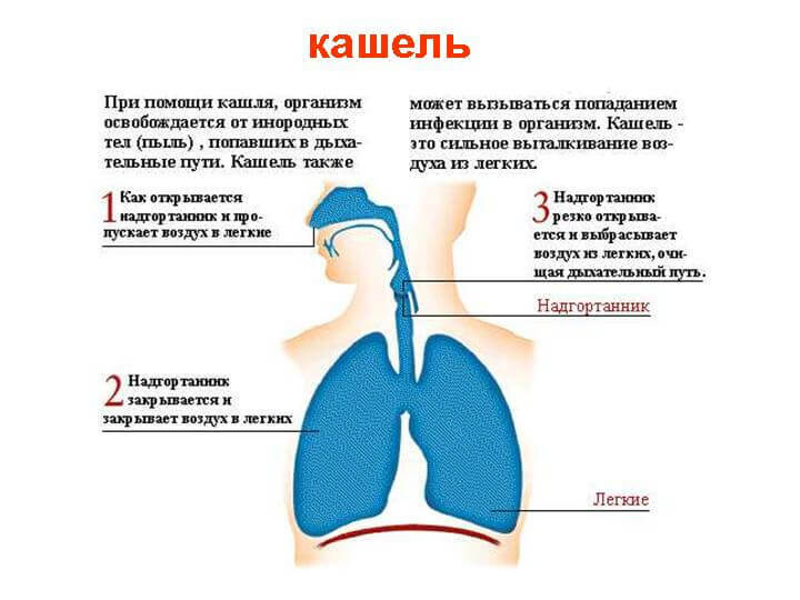
The disease is accompanied by a coughing fit. There is a stabbing pain in a part of the patient's lung, which eventually turns into aching. This is followed by cyanosis of the face (bluish skin color due to the accumulation of carbon dioxide in the blood), pallor. The pain may worsen with movement, breathing, and coughing. The patient may have panic attacks. Trying to relieve shortness of breath and pain, the patient often lies on the sore side or sits down with an inclination to the sore side.
Read also:Fluid in the lungs
Traumatic syndrome affects the general condition of the patient. There is a decrease in blood pressure, severe shortness of breath, the skin becomes cyanotic, the pulse becomes more frequent, and acute heart failure. On exhalation, frothy blood is released from the wound.
During the period of traumatic pneumothorax, air can collect in the subcutaneous tissue of the sternum, the entire neck, face and mediastinum. Feeling at the bulge gives a crunching sensation under the fingers.
Valvular pneumothorax is an extremely serious condition of the patient. There is a penetration of the entire air mass into the pleural area, the exit of which is impossible back. Symptoms and disorders are extremely pronounced. Shortness of breath, sometimes loss of consciousness, cyanosis increases rapidly. Sharp and stabbing pain that can radiate to the scapula, shoulder and abdominal cavity.
In severe condition, the veins of the upper extremities and cervical veins may swell. The affected side is enlarged due to the widening of the gaps between the ribs. Rising heart rate and lowering blood pressure.
Subcutaneous emphysema progresses, speech is inhibited. With its growth, in the wrong place, it often causes cardiac and pulmonary insufficiency.
The child's appearance occurs when the lungs are not properly expanded. In a child under the age of three, this can develop into pneumonia.
Symptoms often do not appear clinically. With complications, signs of pneumothorax in children are:
- body cramps;
- pallor;
- increased heart rate;
- temporary cessation of breathing.
The diagnosis is made by examination. Most often, a chest x-ray or CT scan (computed tomography) is needed.
Diagnostics

Examination is done by auscultatory method (listening with a stethoscope). Thus, weakening or complete absence of breathing in a part of the diseased lung is revealed.
By tapping (percussion), the doctor hears a loud and low sound.
During a prophylactic fluorography scan, a specialist may suspect a mantle-like type of pneumothorax. In order to make sure of this, he prescribes X-ray diagnostics.
Another examination method is an X-ray. With a disease, the picture shows the causes and symptoms of its manifestation. There is a lumen with no pattern of the lung, this is due to the fact that air collects in the pleural cavity. The diaphragm can move downward.
The mediastinum leaves, in the direction of a normally functioning lung. Atelectasis of the lung may form (partial or complete contraction of the lung tissue, which leads to a decrease in the amount of air in the lungs and disrupts ventilation of the alveoli).
The posterior side view will show a thin line of the visceral pleura (no more than 1 mm). The enlightenment line is shown by the lateral position.
Thoracoscopy is used for diagnosis. Thanks to her, the pleural cavity of a sick person is examined. A special device - a tarakoscope - is introduced through a hole made in the chest wall. With its help, gas and an increase in pressure inside the pleura are detected.
Read also:Pulmonary pleurisy: what is it, causes, symptoms and treatment
Computed tomography is one of the best methods of examination in this area. It makes it possible to detect the presence of gas in the pleura. This examination can reveal the probable origin of a spontaneous pneumothorax that is not recognized on x-ray.
Complications
This disease, with illiterate treatment, can lead to some complications. The most common are:
- with a valve form, subcutaneous and mediastyl emphysema may develop;
- a tear in the lung tissue can provoke bleeding inside the pleura;
- the formation of adhesions that interfere with the expansion of the lung. Because of them, serous-fibrinous pleurisy develops;
- accumulation of pus in the pleural cavity (pleural empyema);
- reventilation pulmonary edema.
A long course of the disease (especially without timely seeking medical help) can cause the replacement of lung tissue with connective tissue. The lungs shrink and lose their elasticity.
Pulmonary heart failure develops, which can lead to death.
Treatment
The mild form, which passes without symptoms of the respiratory system, sometimes does not require quick hospitalization and even treatment. However, it must be observed by X-ray examination.
With simple forms of its manifestation, the subpleural bladder or bulla is reduced. A defect in the visceral pleura becomes covered with a fluid made up of white blood cells (fibrous effusion).
Then it seals itself and heals on its own. Resorption of all air occurs within 3 months.
Relapses occur in up to 50% of people.
Medication (medications)

The first medical aid is provided by the introduction of such drugs:
- Analgesics - analgin. For excruciating pain, narcotic substances (morphine, omnopon) are injected;
- Antibiotics - tetracycline group (doxycycline, tetracycline and others);
- Tetanus serum.
After surgery, drugs are prescribed blood thinners (heparin, warfarin, etc.).
In case of circulatory disorders, caffeine and camphor are administered.
Preventive methods of recurrent type use the method of chemical pleurodesis. Injecting irritating agents:
- magnesium silicate;
- glucose;
- silver nitrate solution.
Surgery

With a penetrating wound into the chest cavity (for example, in conditions of hostilities), after which it develops pneumothorax and unilateral air leakage occurs, there is a need for a first-aid interference.
For this, decompression needles have been developed, which, with correct manipulations, pump out the air entering the pleural cavity, due to which the pressure can be stabilized.
Also, special occlusive dressings (films) have been developed, on an adhesive basis, which adhere even to wet skin, creating an airtight seal at the wound site and not allowing the pressure in the chest to equalize atmospheric.
Pneumothorax in any of its manifestations requires surgical intervention. These include the following types of procedures:
- Closed type - with the help of puncture, air is pumped out of the pleural cavity.
- Open type - thoracoscopy or thoracotomy is performed to check the lung tissue and pleura. The defect is sutured, thereby stopping the flow of air into the pleural cavity. Then the event is repeated as in the closed type.
- Valvular pneumothorax - puncture is performed using a thick needle. Thereafter, they are treated surgically.
- Recurrent pneumothorax - remove its causes surgically. Often, a drainage tube is installed to evacuate air, rather than an ordinary pleural puncture.
Read also:Diffuse pulmonary fibrosis: what is it, causes, symptoms, how to treat, prevention and prognosis
Postoperative exercise
After injury or any other injury that entailed pneumothorax, it is necessary to restore your physical form. For this, exercise therapy exercises are used, which begin through 3-4 weeks after being wounded.
It is necessary to start (as with any workout) with light, gentle exercises, gradually increasing the load. The most common exercises are breathing exercises (inflating balls, breathing into a tube). Strelnikova breathing exercises are actively recommended by doctors.
It is recommended to go to the pool and do race walking.
Complementary and alternative home treatments
Self-treatment for this disease is impossible - seeking help from a qualified specialist is the only right decision. But you can combine medication with traditional medicine recipes.
Herbal treatment

Recipe from Veronica officinalis. To brew 1 dining room spoon of crushed plant in two glasses of water. Cover and leave for 2 hours. Filter before use. Drink infusion 1 teaspoon 4 times per day. After several receptions of the infusion, the patient's appetite increases markedly.
Cloudberry juice is very useful. Drink it instead of tea, several times a day.
To restore strength, a knotweed decoction is used. It lowers blood pressure and increases blood clotting.
For the broth, take 1 tablespoon herbal raw materials, pour 250 g of boiled water over it. Then simmer in a water bath for 15 minutes. Then remove from heat and cover for 2 hours. Before use, filter and drink 1 tablespoon 3 times a day
Prophylaxis
It is recommended to observe the following precautions:
- avoid barometric pressure drops (flights in airplanes that are not equipped with atmospheric pressure stabilizers, exclude deep diving and rock climbing);
- quit smoking;
- for 3 months, suspend sports and do not lift weights.
Forecast
Usually, uncomplicated manifestations of the disease do not have adverse consequences for the human body. The prognosis is determined by the degree and size of the lesion of the respiratory system. The sooner help is provided, the less likely the condition will worsen.
Up to 40% of people may experience a relapse. Usually recurrence occurs within six months after the first attack.
Death rate:
- HIV-infected - no more than 25%.
- In people with congenital cystic fibrosis, with the development of unilateral pneumothorax 5%. Double sided gives 25%.
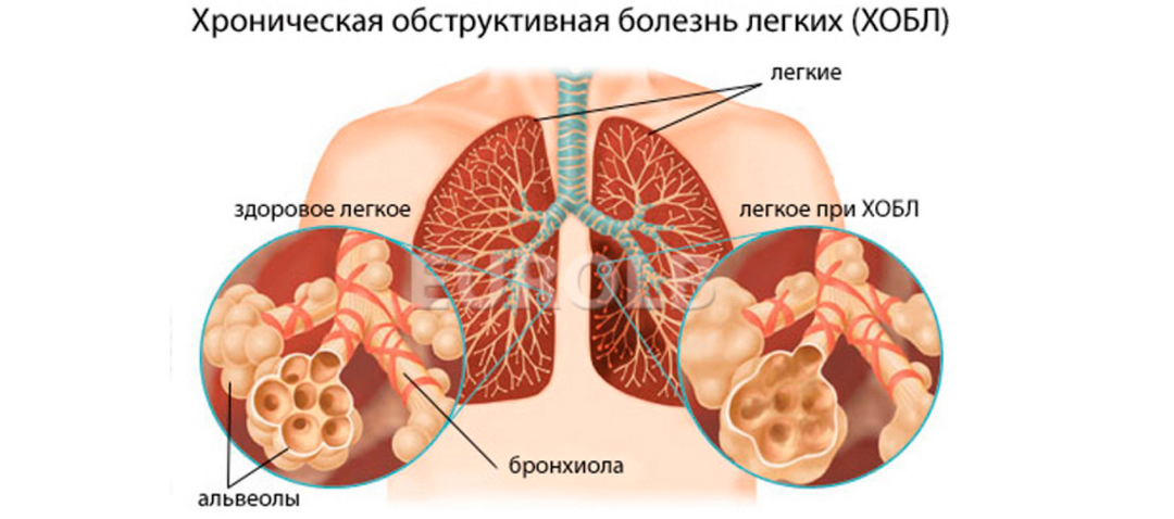
- People with chronic obstructive pulmonary disease on average 5%.