Arthritis: what is it, causes, symptoms, treatment, prognosis
Content
- What is arthritis?
- Sign and symptoms
- Causes and risk factors
- Epidemiology
- Pathophysiology
- Diagnostics
- Treatment
- Forecast
What is arthritis?
Arthritis Is an acute or chronic joint inflammation that is often accompanied by pain and structural damage. Arthritis is not synonymous with arthralgia, which refers to pain localized to a joint regardless of the origin of the pain (which may or may not be related to joint inflammation). Arthritis affected both Neanderthals and ancient Egyptians, but it wasn't until 1886 that Dr.John C. Spencer coined the term "osteoarthritis". More than 100 different types of arthritis have been described, the most common of which is osteoarthritis or degenerative arthritis, which is non-inflammatory arthritis. Inflammatory arthritis can occur in several conditions, and inflammation can be caused by autoimmune processes (rheumatoid arthritis, psoriatic arthritis, ankylosing spondylitis etc.), inflammation caused by the deposition of crystals (gout, pseudogout, underlying calcium phosphate disease) or infections (
septic arthritis, Lyme arthritis). Inflammatory arthritis may also accompany others autoimmune diseases connective tissue such as systemic lupus erythematosus, Sjogren's syndrome, scleroderma, myositis, inflammatory bowel disease, celiac disease, etc.Sign and symptoms
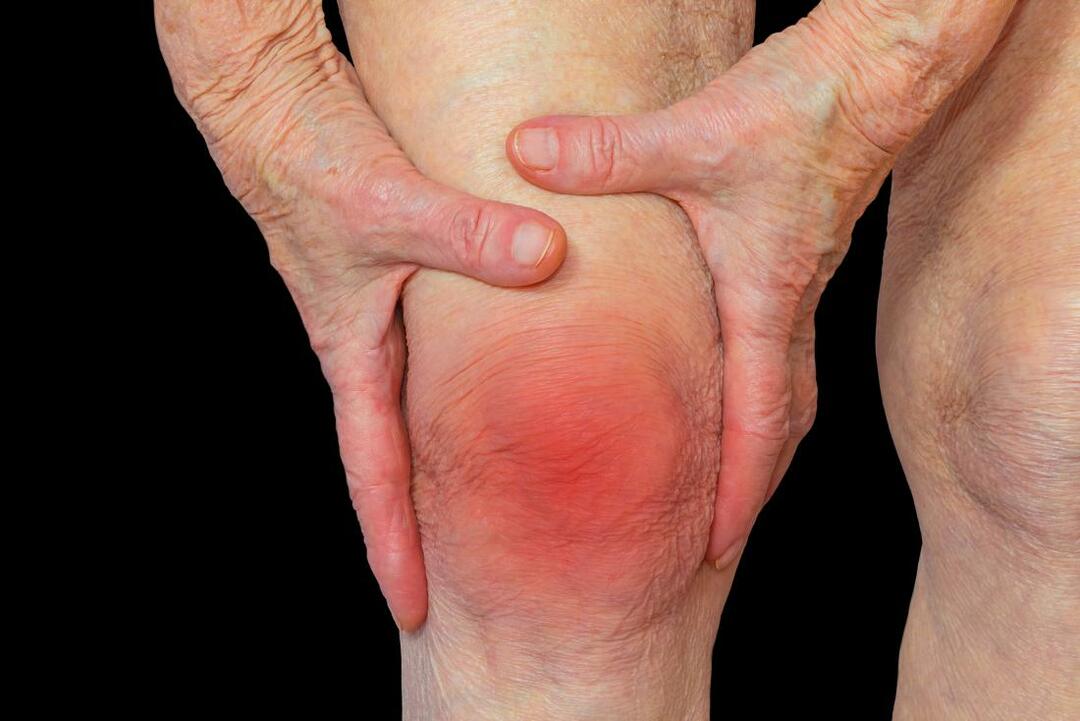
Pain, which can vary in severity, is a common symptom of almost all types of arthritis. Other symptoms include swelling, joint stiffness, and pain around the joint (s). Arthritic diseases such as lupus and rheumatoid arthritis can affect other organs in the body, causing many symptoms. Symptoms and signs may include:
- inability to use a hand or walk;
- Stiffness that may worsen in the morning or after activity
- malaise and fatigue;
- weight loss;
- poor sleep;
- muscle pain;
- difficulty moving the joint.
With advanced arthritis, significant secondary changes often occur. For example, symptoms of arthritis can make it difficult for a person to move around and / or exercise, which can lead to side effects such as:
- muscle weakness;
- loss of flexibility;
- decreased aerobic fitness.
These changes, in addition to the underlying symptoms, can have a huge impact on quality of life.
Causes and risk factors
The etiology of arthritis depends on the type of arthritis. The main factors contributing to osteoarthritis are old age, female gender, joint injuries, and obesity. Several genetic factors have been described, such as mutations in genes encoding collagen types II, IV, V and VI.
Rheumatoid arthritis (RA), on the other hand, is an autoimmune systemic inflammatory disease. The interaction between several genetic factors (HLADRB1 and others) and environmental factors (smoking) leads to activation and dysfunction of the immune system, which leads to inflammation in RA.
In gout, prolonged hyperuricemia leads to the deposition of uric acid in the joints, which then leads to their inflammation. There are several genetic mutations that can cause hyperuricemia, although it accounts for less than 10% of cases of gout. Most patients with gout are under-excreted. they cannot get rid of all the uric acid that is produced in them as a result of endogenous or exogenous purine metabolism. Male gender, old age, chronic kidney disease, alcoholism and certain medications such as diuretics are additional risk factors for hyperuricemia and gout.
Septic arthritis is an acute arthritis that rarely occurs in the general population, but patients with pre-existing risk factors such as immunodeficiency, advanced age, diabetes, prosthetic joints, rheumatoid arthritis and intravenous drug abuse are at higher risk.
Arthritis is often seen in patients with other autoimmune diseases and is one of the most common clinical manifestations in patients with systemic lupus erythematosus (SLE). Other conditions often associated with arthritis include inflammatory bowel disease, psoriasis, celiac disease, Sjogren's syndrome, systemic sclerosis, dermatomyositis, mixed connective tissue disease (MCTD), etc.
Epidemiology
More than one third of the population suffers from imaging arthritis, and this number is bound to increase with the average age of the population. Osteoarthritis is the most common arthritis. From 19% to 30% of adults over the age of 45 suffer from osteoarthritis of the knee joint, 27% suffer from osteoarthritis of the hand and 27% from osteoarthritis of the hip. It is estimated that 40% of men and 47% of women will develop osteoarthritis during their lifetime, with the incidence rising to 60% if they have a BMI greater than 30.
The incidence of gout is more than 45 cases per 100,000 people. It is noteworthy that both the incidence and prevalence of gout has more than doubled over the past few decades. The prevalence of pseudogout in the adult population ranges from 4% to 7%, with more than half of patients suffering from knee arthritis.
Read also:Wegener's granulomatosis (granulomatosis with polyangiitis)
Rheumatoid arthritis occurs in about 1% of Caucasians, and women suffer more often than men (the lifetime risk is 3.6% in women versus 1.7% in men). The disease usually occurs in early adulthood, with a prevalence of 5% in women over 65.
Septic arthritis is usually caused by bacterial culture in a joint already affected by arthritis through hematogenous spread, most often as a result of a skin or urinary tract infection. Septic arthritis has a prevalence of 0.01% in the general population and 0.7% in patients with rheumatoid arthritis.
Pathophysiology
Osteoarthritis is characterized by a degenerative cascade of progressive loss of cartilage that results in bone damage. Common signs include subchondral cysts, osteophytes and thickening of the subchondral plate. Interleukin-6, monokines, interferon-induced protein-10 and chemotactic protein of macrophages induce proteolytic enzymes such as matrix metalloproteinases, serine proteases and cysteine proteinases, and lead to degradation of the articular collage. Calcification of the surrounding articular cartilage reduces thickness and eventually destroys the cartilage matrix. Old age is also associated with decreased chondrocyte function, increasing the susceptibility to osteoarthritis.
The symptoms of rheumatoid arthritis are usually more severe than the symptoms of osteoarthritis. Rheumatoid arthritis Is a systemic and chronic inflammatory condition caused by an autoimmune response to an environmental trigger. Degradation of cartilage and ultimately bone is preceded by activation of endothelial cells and hyperplasia of synovial cells. Pathology occurs after aberrant production of inflammatory mediators (such as alpha tumor necrosis, interleukins 1, 6 and 8, and others after exposure to an antigenic pathogen).
The monosodium salts of gout precipitate in the form of needle-like crystals. This crystallization is more likely in colder parts of the body and in acidic environments. Destabilization of these deposited intra-articular uric acid crystals leads to an inflammatory response mediated by IL-1, resulting in the typical exacerbation of gouty arthritis. This process differs in pseudogout, when inorganic pyrophosphate from chondrocytes combines with calcium to form calcium pyrophosphate dihydrate. This crystal is deposited in the joint spaces prone to osteoarthritis. The damage caused by crystals in pseudogout includes fragmentation of bones and cartilage, as well as the formation of osteophytes and subchondral cysts. Metabolic disorders such as hemochromatosis, hyperparathyroidism, or hypomagnesemia increase the likelihood of calcium pyrophosphate deposition.
Septic arthritis usually an inflammatory response to a monobacterial infection. The ingress of bacteria into the synovial fluid causes the release of cytokines, chemokines and proteases, which destroy cartilage and cause synovial membrane hyperplasia. Toxins produced by bacteria play an additional destructive role in the joint space itself. In adults Staphylococcus aureus is the most common pathogen (strains of streptococci are often found). Infection with gram-negative bacteria is more likely to occur as a result of trauma, intravenous drug use, immunosuppression, or in the elderly or very young.
Diagnostics
History and physical examination are critical in assessing arthritis and determining the type of arthritis, and in differentiating symptoms from non-articular etiology. The first stage of medical examination of a patient with complaints of the musculoskeletal system should be to determine and confirm whether the pain is articular or not. Non-articular pain can be secondary to several pathologies, including fibromyalgia, in which patients experience pain in both articular and extra-articular areas, but at the same time there is no pain in the joints effusion, edema, warmth, or erythema. Tendinitis can also cause periarticular pain, physical examination in these cases usually reveals tenderness along the tendon course without any focal tenderness or loss of range joint movement.
Common symptoms of arthritis are pain, swelling, loss of function, stiffness, deformity, weakness, and instability. They can also be accompanied by fatigue, sleep disturbances, emotional instability, and symptoms of an underlying systemic illness. Arthritis pain usually worsens with activity and late in the day. Inflammatory arthritis also causes pain in the morning and at rest, which may initially be relieved by physical activity, but subsequently increases with prolonged use and physical activity. Patients with fibromyalgia and myofascial pain syndrome usually complain of pain spread throughout the body. Neuropathic pain may be accompanied by paresthesias in the nervous section. Morning stiffness lasting more than 45 minutes is usually associated with inflammatory arthritis, but this is nonspecific as patients with osteoarthritis or non-articular syndromes such as fibromyalgiamay also have prolonged morning stiffness.
Read also:Shoulder-scapular periarthritis
The physical examination is the most important tool in assessing arthritis and arthralgia. Inflammatory arthritis is associated with soreness, swelling, effusion, erythema, and warmth. These features are more evident in acute inflammatory arthritis, but may be less pronounced in chronic inflammatory arthritis. Osteoarthritis can also be associated with soreness, swelling, and effusion (effusion), although erythema and warmth are usually absent. Decreased range of motion and overt joint deformity can also be seen in arthritis.
- Analyzes and visualization.
Laboratory tests and radiographic evaluation can help diagnose and assess the severity of arthritis.
Inflammatory arthritis is associated with increased markers of inflammation (ESR and CRB). Anemia with chronic diseases - a common thing. Leukocytosis can be observed with septic arthritis, as well as with gout, pseudogout and Still's disease in adults, while leukopenia and thrombocytopenia are observed in RA and arthritis associated with SLE. In patients with gout, serum uric acid levels may be elevated, although this should not be the only diagnostic criterion. Serologic tests such as rheumatoid factor, anti-citrullinated peptide antibodies, antinuclear antibodies and more specific autoantibodies should be administered when indicated to aid in diagnostics.
Plain radiographs should be the first imaging modality. Joint space narrowing, osteophytes, and effusion are common manifestations of osteoarthritis. Periarticular osteopenia is the first radiographic sign of inflammatory arthritis and erosions, joint space narrowing, and secondary osteoarthritis that develops later in the disease. Central erosions (gullwing symptom) are a sign of erosive osteoarthritis, while periarticular erosions are seen in RA. Calcifications of entheses can be seen in seronegative spondyloarthritis, especially in psoriatic arthritis and ankylosing spondylitis. X-rays for axial spondyloarthropathies are normal early in the disease, but later may show fusion of the spine (symptom of "bamboo stick") and sacroiliac joint. Axial osteoarthritis, on the other hand, is manifested by osteophytes, disc bulges, and narrowing of the joint space on x-rays. Pseudogout has characteristic radiographic features of chondrocalcinosis, which is calcification cartilage that can be seen on menisci, triangular fibrous cartilage of the wrist, or cartilage of the 2nd / 3rd metacarpophalangeal joint of the hands. Radiographs for gout are normal at onset but may later show hard tophi, periarticular osteopenia, and classic juxtaarticular erosions with overhanging margins.
If the radiographs are not diagnostic, additional testing may be considered. MRI is a very useful tool that can help in assessing the presence or absence of synovitis, erosions, sacroiliitis with much greater sensitivity than x-rays. In addition, MRI can help assess soft tissue or ligament injuries and tumors that are otherwise difficult to identify. CT scans can help detect bone deformities and detect chondrocalcinosis, but is mostly done if MRI is not possible. Musculoskeletal ultrasound, which is an evolving technique, is extremely useful, especially in assessing peripheral arthritis, and can help in assessing the presence of or absence of synovitis, effusion, erosion, structural defect such as rotator cuff or meniscus rupture, and may also aid in performing ultrasound aspiration / injection. Nuclear medicine joint scans are rarely indicated due to their high sensitivity but low specificity, but can help in assessing the presence or absence of inflammation, especially for evaluating prosthetic infections joints. Dual-energy computed tomography is another emerging X-ray technique that is highly accurate in helping diagnose gout.
The study of synovial fluid is one of the most important tests, especially for the initial diagnosis of arthritis. Cell counting and differentiation, crystal evaluation under polarized light microscopy, bacterial / acid-fast bacilli / fungal cultures and Lyme DNA PCR should be performed on synovial liquids if necessary. Degenerative arthritis is usually associated with a cell count of less than 2000 cells / mm3, whereas in inflammatory arthritis, the cell count usually exceeds 5000 cells / mm3 and can reach 50,000 cells / mm3. More than 50,000 cells / mm3 and / or more than 90% of neutrophils in synovial fluid analysis should cause suspicion of septic arthritis, although this can also be seen in acute gout or pseudogout. Gout crystals are needle-shaped and have strong negative birefringence. Pseudogout crystals are diamond-shaped and exhibit weekly positive birefringence. Basic calcium phosphate crystals are not visible under polarized light microscopy and require special staining to confirm positivity. Synovial biopsy is rare but may be considered, especially in cases of monoarthritis where other methods have failed to establish a diagnosis.
Read also:The initial stage of diabetic foot: causes, symptoms and treatment
- Differential diagnosis.
Arthritis should be differentiated from non-articular pain, including pain caused by fibromyalgia, myofascial pain syndrome, neuropathy, tendinitis and complex regional pain syndrome. Physical examination, as well as laboratory and x-ray examinations, can help in this distinction.
There are over 100 different types of arthritis described, and it is important to establish an accurate diagnosis before starting treatment.
Treatment
Treatment for joints suffering from osteoarthritis should aim to reduce pain and improve function. Typically, optimal treatment requires a combination of non-drug (or conservative) and pharmacological treatments.
Non-drug treatments include special exercises, physical therapy, bracing, acupuncture, and weight loss. Topical and oral medications are used in the pharmacological treatment of osteoarthritis. Commonly used medications include oral and topical non-steroidal anti-inflammatory drugs (NSAIDs), topical capsaicin, and duloxetine. Corticosteroids can be injected directly into the joint. In most cases, topical NSAIDs, capsaicin, and other ointments are first-line therapy, and oral NSAIDs should be started if the methods described above do not relieve symptoms or the disease is more systemic character. Duloxetine may be useful in patients who have medical contraindications for NSAIDs and have been shown to be beneficial, especially for osteoarthritis of the knees.
If both non-pharmacological and pharmacological treatments fail, intra-articular corticosteroid injections may relieve symptoms. The use of opioids should be avoided. Surgical replacement of the affected joint (s) is used for refractory symptoms and can be very effective. Patients may experience postoperative complications, and limited function in the immediate postoperative period is not uncommon. Postoperative physiotherapy is essential to improve patient outcomes.
The emphasis in the pharmacological treatment of rheumatoid arthritis and seronegative spondyloarthropathies is on early remission of the disease and prevention of radiographic progression. Early use of disease-modifying antirheumatic drugs (DMARDs) and biologics is more effective than treatment with glucocorticoids and NSAIDs. Anti-inflammatories can be used as an adjunct to reduce inflammation while the disease remains active. Regular monitoring of symptoms and dose adjustments continues until symptoms are in remission. In severe cases, corticosteroids may be needed to treat flare-ups of the disease.
With gouty arthritis, flare-ups can be very debilitating and painful. The use of anti-inflammatory drugs can provide significant relief and should ideally be started within 24 hours of a gout flare-up. These include oral corticosteroids, NSAIDs such as high-dose indomethacin or naproxen, or colchicine. The use of intra-articular corticosteroid injections may be beneficial in a patient with a small-joint lesion, whereas the use of intramuscular or intravenous corticosteroids may be used if the patient is unable to take the medication orally. Medications that lower uric acid levels do not play a useful role in treating an exacerbation, but are recommended for patients with recurrent exacerbations. chronic kidney disease, nephrolithiasis or tofus. The goal is to reduce serum uric acid, which then lowers the content of uric acid inside the joint, which ultimately leads to the disappearance of symptoms gout.
Treatment for septic arthritis requires drainage of the affected joint and the use of antibiotics. Cultures and sensitivity of the joint fluid determine the use of antibiotics in the joints.
Forecast
Osteoarthritis is an incurable, progressive disease. The prognosis depends on the number of joints affected and the severity of the disease. Rapid progression is likely in elderly patients, those with obese, varus deformity and multiple joint lesions. After joint replacement, the results are good, but there is no permanent prosthesis; thus, repeated interventions may be required after 10-15 years.
With recent advances in therapy, the prognosis of rheumatoid arthritis has improved significantly, although the incidence and mortality in rheumatoid arthritis is significantly higher than in the general population, mainly due to extra-articular manifestations.