Wirsung's duct is not visualized: what does it mean? Find out from our article!
The most informative diagnostic technique for diseases of the pancreas is ultrasound. It allows you to assess the state of all structures and parts of the organ, including the Wirsung duct. The main criteria by which the doctor determines the diseases and abnormalities of the pancreas are echogenicity (the ability to reflect sound), the size and appearance of the organ. Sometimes in the ultrasound protocol you can find a phrase that causes concern for patients: "the Wirsung duct is not visualized." What does such a conclusion mean, and what pathologies or ailments can it indicate?

Wirsung's duct is not visualized: what does it mean?
Content
- 1 What is the Wirsung duct?
- 2 Indications for ultrasound of the pancreas
- 3 Preparation for ultrasound examination of the pancreas
-
4 Pancreatic ultrasound: norms and pathologies
- 4.1 Video - Anatomy of the Pancreas
- 5 Virsungov duct on ultrasound
What is the Wirsung duct?
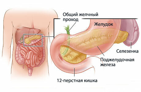
Wirsung (pancreatic) duct is the main highway of the pancreas, into which small, secondary ducts flow. It stretches from the tail of the organ through its entire body, reaches the back, and is about 20 cm long, and the width differs in depending on the site - at the beginning about 2 mm, then 2-3 mm, and at the end, where the sphincter of Oddi is located, the diameter can reach 4 mm. The shape of the canal almost exactly repeats the shape of the pancreas, but sometimes it can be annular or S-shaped.
The structure of the Wirsung duct depends on the characteristic features of the human body - in most people it connects with the common bile duct (common bile duct) and ends in the duodenum.
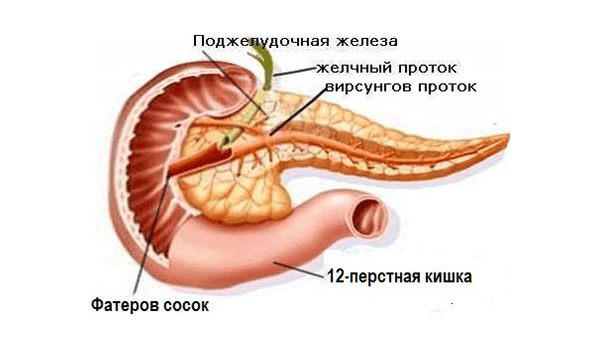
Location of the Wirsung duct
There are four types of merge data backbones:
- the choledoch and the Wirsung duct flow into the duodenum, forming something like an ampoule, and the intestinal sphincter, when contracted, completely closes them (found in 55% of people);
- the ducts are connected next to the duodenum, but do not merge together (observed in 33% of cases);
- the lines merge at some distance from the intestine, but end in it and are in contact with the walls (8.5% of cases);
- ducts flow into the intestine independently of each other (4%).
Along the entire length of the channel, additional ducts of a smaller size and diameter flow into it. Their number also differs depending on the structure of the digestive system: with the main type of the gastrointestinal tract, a person has 20-35 secondary ducts, with a loose one - about 60. In the first case, the small channels are located 1.5 cm apart, but in the second, they are much smaller.

Virsungov channel
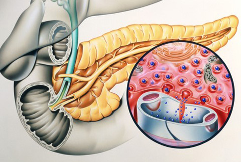
The main function of the duct is to transport pancreatic secretions, which take part in the digestion process, to the gastrointestinal tract. Pathologies of this part of the organ speak of various diseases associated with the work of the pancreas, and are also an important criterion for ultrasound diagnostics.
Indications for ultrasound of the pancreas
The indications for ultrasound examination of the pancreas include:
- pain localized in the left hypochondrium or radiating to this area, which lasts for several weeks;
- discomfort, heaviness or heartburn that occurs even after taking a small amount of food;
- the appearance of a yellow tint to the skin and mucous membranes;
- dysfunction of the digestive system, accompanied by constipation or diarrhea.

Pancreas ultrasound
When any of the above symptoms appear, the patient needs a comprehensive diagnosis of all organs involved in the digestion process, including the pancreas.
Important! The work of the pancreas is closely related to the activity of the liver, therefore, in case of any unpleasant sensations in the area of the liver, it is necessary to check the pancreas as well.
Preparation for ultrasound examination of the pancreas
As a rule, ultrasound examination does not require special preparation from the patient, but the pancreas anatomically located next to the hollow organs (stomach, intestines), therefore, the diagnostic results can be distorted by the from there air. For inaccuracies in the ultrasound scan of the pancreas, the patient must properly prepare for the procedure.

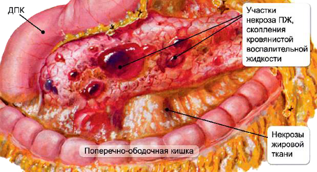
This is what an inflamed pancreas looks like.
The research is best done in the morning - at this time, the indicators will be the most informative. It is recommended to refuse food intake within 12 hours before the diagnosis, and to exclude a few days before consumption of carbonated drinks, muffins, fresh bread, legumes and other foods that can cause gas formation. If an ultrasound scan is performed without prior preparation, the accuracy of the results may decrease by 40%.
Pancreatic ultrasound: norms and pathologies
When conducting an ultrasound scan, the doctor examines the pancreas with a special sensor, and an image appears on the screen by which one can judge its condition. There are several indicators that allow you to determine the norms and pathologies in the structure of the organ.
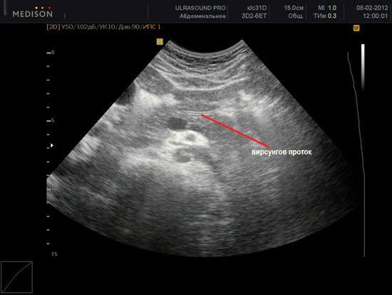
Virsungov duct on ultrasound
- In a healthy person, the body of the pancreas has a homogeneous structure (minor inclusions are allowed no more than 3 mm in size), clear and even contours, located in the center of the spinal column exactly under stomach.
- The brightness and intensity of the picture on the monitor depends on the echogenicity of the organ, that is, the ability of its tissues to reflect sound waves - normally the echogenicity of the pancreas is the same as that of the spleen and liver.
- The organ on ultrasound should be well visualized - so that the doctor can determine the size of all its parts. The width of the body in the absence of pathologies is 21-25 mm, the head is 32-35 mm, the tail is 30-35 mm.
To assess the large vessels that are close to the pancreas and supply it with blood, duplex scanning of the organ is additionally performed. The interpretation of the diagnostic results is carried out taking into account all indicators and is carried out exclusively by the attending physician.
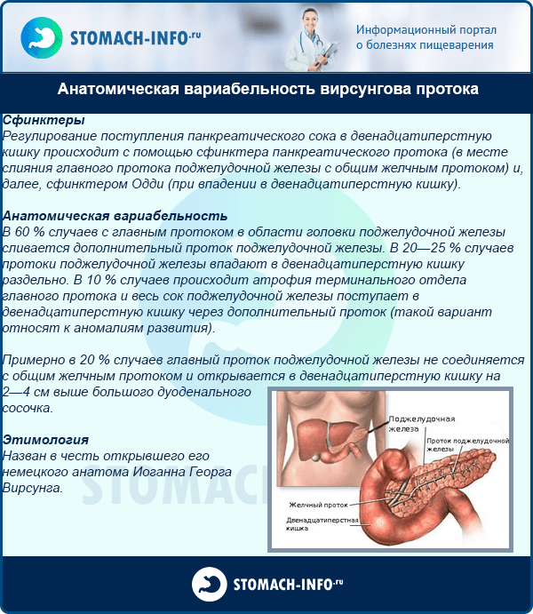
Anatomical variability of the Wirsung duct
With pancreatitis, tumor processes and other diseases of the pancreas, the contours of the organ become blurred, uneven, it increases in size, and the echogenicity increases significantly, or, conversely, decreases. Sometimes changes are observed in the entire organ, and sometimes - in its individual segments.
For reference! The size of the pancreas largely depends on the individual characteristics of the organism, and can vary significantly from person to person. When making a diagnosis, as a rule, the upper limits of the norm are taken into account, but in the absence of serious changes in the structure of tissues and biochemical blood tests, their excess does not indicate the presence pathologies.
Video - Anatomy of the Pancreas
Virsungov duct on ultrasound
Changes in the Wirsung duct are one of the most informative diagnostic criteria in determining pancreatic diseases. Normally, it is well visualized on the monitor, has a width of about 2 mm and an even shape, which depends on the characteristics of the organism.
It should be noted that the phrase "Wirsung's duct is not visualized" in the ultrasound protocol does not always speak of work pathologies pancreas - sometimes this can be due to the characteristic structure of the organ and its deep location in the body person. According to medical statistics, it is possible to clearly visualize the Wirsung duct only in 56-80% of cases - in other people it is impossible. But changes in the contours of the canal, its expansion or narrowing indicates the presence of diseases that require immediate treatment.

Ultrasound of the pancreas
| Disease | Changes in the Wirsung duct |
|---|---|
| Pancreatitis | Expansion of the canal along its entire length or in some areas, thickening of the walls, uneven contours, the presence of calcifications |
| Cystosis | The appearance of areas of expansion and narrowing, the presence of cysts and calcifications |
| Giardiasis | Thickening of the canal walls |
| Tumor diseases | Blockage or narrowing of the duct in one place |
| Cystic fibrosis | Multiple sites of constriction, blockage in the region of the head of the organ, formation of stones |
Sometimes the functions of the pancreatic duct are impaired due to congenital anomalies of its structure - bifurcation, splitting, etc.

Pancreatic tumor
Important! If it is impossible to visualize the Wirsung duct on an ultrasound scan, and it is necessary to assess its condition and work in order to make an accurate diagnosis, patients are prescribed additional research methods - endoscopic retrograde cholangiopancreatography, sonography, computer tomography.
The state of the Wirsung duct is one of the most effective diagnostic criteria that allows you to identify abnormalities in the work of the pancreas. With any changes in this part of the organ, the patient needs a comprehensive diagnosis and competently prescribed treatment, otherwise the dysfunction of the digestive system can lead to serious consequences.