Imperfect osteogenesis
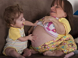 Imperfect osteogenesis is a congenital disease of bones, as well as individual connective tissue structures. Another name for imperfect osteogenesis is a disease of fragile bones. The disease is transmitted by inheritance, i.e. Is genetically determined.
Imperfect osteogenesis occurs in both men and women, the incidence rate is 1 newborn per 12 000-15 000 children. This pathology was first mentioned in the 17th century. In imperfect osteogenesis, growth, skin lesions, muscle tissue, blood vessels, teeth, tendons and hearing organs are disturbed.
Despite the difficulties faced by people with imperfect osteogenesis, most patients lead a full-fledged lifestyle. At the moment, the treatment of imperfect osteogenesis is becoming more and more popular. Nevertheless, it is still impossible to completely cure a patient with such a disease.
Imperfect osteogenesis of the cause of
Imperfect osteogenesis refers to heterogeneous hereditary diseases affecting the connective tissue. The disease is described by osteopenia and some other clinical signs of osteogenesis.
The main cause of this pathology development is a hereditary mutation of collagen genes. Rare is the spontaneous occurrence of mutations in the genes of this protein. The consequence of gene mutation is a violation of collagen synthesis, hence the pathologies in the formation of bone and cartilaginous tissues.
Insufficient amount of synthesized collagen( not mutated) is also one of the reasons for the development of imperfect osteogenesis. In this case, the disease proceeds in a more mild form. Imperfect osteogenesis of this nature is expressed in individual fractures of the limbs, after the puberty, the number of fractures, as a rule, decreases.
Modern medicine distinguishes several types of imperfect osteogenesis, taking into account radiographic, clinical, collagen proteogenic changes.
Imperfect osteogenesis types:
Type I - a weakly expressed form, a dominant type of inheritance. Imperfect osteogenesis, characterized by brittle bones, the presence of blue sclera.
Type II - perinatal-lethal.
Type III - deformation of the skeleton is progressive.
Type IV - the dominant type of inheritance, the deformations of the skeleton are not pronounced sharply, sclera are normal.
Many believe that imperfect osteogenesis develops against the background of qualitative and quantitative violations of collagen type I synthesis. Imperfect osteogenesis of type I is characterized by a reduced level of synthesis of normal collagen. Type II, like type IV imperfect osteogenesis, is caused by a decrease in the total amount of collagen due to reduced stability, although the collagen synthesis process passes without any disturbances.
Imperfect osteogenesis symptoms
Each type of imperfect osteogenesis has its characteristic symptomatology.
So, I type of imperfect osteogenesis in children is manifested by osteoporosis, as well as frequent fractures of bones. After a 10-year-old age, the incidence of fractures decreases, but again rises after 40 years. The blue sclera and the early appearance of the senile rim are noted. Patients( about 50% of cases) with this type of imperfect osteoporosis of low growth, some of dentin remains normal, some patients have dentine opal. In type I, aortic changes occur in a number of cases, there is mitral defect( in 20% of cases, mitral valve prolapse occurs), nasal bleeding.
Type II of imperfect osteogenesis is characterized by fetal death before birth or early neonatal death. Fractures are multiple and frequent, easily occur. There are 3 groups:
Group A. Damage to the head, limbs in the fetus are noted during pregnancy due to the fragility of connective tissue formations. The brain area of the skull is enlarged, the thorax, on the contrary, is reduced, the limbs are short and curved. There are severe cases of calcification of the aortic walls and endocardium. Such children are born very small growth( about 25-30 cm).
Births before the term, with 20% of such premature births - stillbirth. Some sick children die in the early days, some live up to the 4th week. Pathological changes can be detected even before the birth of the child with the help of X-ray study: the femurs are broad, there are wavy edges, ribs with bunions, the thorax is short. When conducting medical genetic counseling in probands, there is a possibility of a molecular defect combined with heterozygousness of mutations localized in the collagen gene. The inheritance is autosomal dominant.
Group B. Phenotypically similar to group A. Nevertheless, disturbances in the respiratory system are not so pronounced, children with imperfect osteogenesis live for several years. There are shortened tubular bones and changes in the structure of the ribs, however, their fractures are rare. Presumably heredity is autosomal recessive.
Group B. Such cases of imperfect osteogenesis are rare. Characterized by stillbornness or death in the first month. In patients, small growth, fineness of tubular bones( in particular diaphysis), skull bones without ossification are noted. Presumably an autosomal recessive type of inheritance.
Patients with type III imperfect osteogenesis are rare. During childbirth, fractures sometimes occur, the body of the newborn is shortened, while the mass may be within normal limits. O-shaped deformation of free extremities, kyphoscoliosis is characteristic. The cause of death of about half of the patients are various changes in the skeleton and circulatory system. Osteoporosis is pronounced, the growth of bones in length is broken along with their ossification. In the growth zones of bone tissue, uneven calcification is observed, which leads to the appearance of spotting( the so-called "corn kernels").Heredity is not exactly known.
IV type of imperfect osteogenesis in children is most often manifested as a disorder in the skeleton. This type has a large variability of osteopenia, the number of bone fractures, age, blueness sclera. Usually with age, the number of bone fractures decreases, bone calluses are formed, after 30 years in some patients with imperfect osteogenesis of this type, hearing is disturbed. According to the state of dentin, patients are divided into 2 groups: some have sharp changes in dentin( opal teeth), others have no changes in dentin.
Imperfect osteogenesis treatment
Diagnosis of imperfect osteogenesis relies, first of all, on the clinical picture of the disease. To establish an accurate diagnosis, a specialist consultation( endocrinologist, orthopedist or geneticist) and the appointment of laboratory tests to exclude other diseases are necessary.
Laboratory methods include molecular analysis of collagen and its biochemical analysis, investigation of skin biopsy material. Instrumental methods of investigation include: X-ray examination( determination of osteopenia, various fractures, bending of bones, vertebral column fractures, intercalary bones in the skull), bone biopsy, densitometry.
The main goal of treatment of imperfect osteogenesis is to reduce the number of fractures in the patient, increase the mineralization of bones to reduce their fragility, increase bone mass. An important point of treatment is the adaptation of the child, as well as of his parents, to the changed lifestyle due to illness. This will avoid in the future fractures of bones.
The entire course of treatment consists of:
- Medication. To combat imperfect osteogenesis, preparations are used: bisphosphonates, growth hormone, vitamin D3, calcium gluconate, magnesium salts and potassium salts, ergocalciferol, complexones, glycerophosphate.
- Operative intervention. It is necessary to eliminate fractures, strengthen bones, correct changes due to deformity, prosthetics. Sometimes surgical treatment is not one operation: because of the growth of bones previously inserted pins require replacement.
In the treatment of fractures, flexible titanium rods with hypoallergenic properties are used. Such rods are well fixed with a fracture, so the need for additional immobilization of the body region disappears. The usual use of gypsum dressings causes difficulties in care, it causes new fractures. It is also possible to use various grafts, bone plastics and other metal structures.
Given the increased fragility of the bones in the patient, in some cases, orthopedists produce osteoclasis to correct the deformation change in the bone, then the limb is fixed with a plaster bandage. Also suitable for the immobilization of body stretching.
In addition to the main methods of treatment of imperfect osteogenesis, also apply: exercise therapy, physiotherapy, osteosynthesis. A course of rehabilitation and psychotherapeutic support is provided for both the patient and family members.
The full course of treatment does not allow to fully cope with such a serious disease as imperfect osteogenesis, therefore, all the above methods allow only partial elimination of symptoms and facilitate a way of life for the patient without affecting the underlying cause of the disease.