Cyst of epididymis
Spermatoceles or cysts of the epididymis occur in the testicle due to obstruction in the duct( seminal) of the appendage of one of the parts. This disease manifests itself more often in boys from 6 to 14 years. Gradually, the cyst increases, adversely affecting the surrounding tissues, which is the cause of compression of the ducts of the epididymis and disruption of the male gonad.
What is a cyst that forms in the epididymis?
A benign oval or rounded formation located in the appendage of the testicle is the cyst of the epididymis of one of the testicles. It forms a cystic formation of a dense fibrous membrane, where a serous fluid has accumulated. A neoplasm arises due to many factors and develops as a result of a disturbance in the outflow of fluid produced by the appendage, which liquefies the sperm. It turns out that the liquid substance is produced, but it is not excreted from the body. As a result, cavity formation takes place.
Why does the spermatoceles appear?
The causes of the formation of cysts( spermatogenic) can be congenital, which occur even in the embryonic period, due to the non-extinction of the Müllerian duct. Also, the testicles can be filled with fluid without sperm.
The causes of sperm cells can be acquired. Occur due to injuries, inflammatory diseases( infection of the seminal ducts).But there are cases that the cyst of the appendage of one of the testicles occurs without any obvious reasons.
What is the danger of cysts in the epididymis?
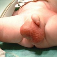
Cystic formation in itself is considered a safe phenomenon, not a threat to human life. But progressive unfavorable changes gradually lead to the fact that the reproductive masculine function is violated. Because of the growth of the cyst in the testicle, the surrounding tissues are squeezed, closing the lumen of the spermatic cord. Because of the progressive growth of the cyst increases, and significantly, the size of the scrotum. As a consequence, it prevents you from sitting, walking, violating urination, sex life.
Diagnosis of the cyst of the epididymis of the
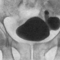
The patient is carefully examined. Determine in him a dense formation( painless) in the head or tail of the epididymis. Rarely, but it happens that the presence of a cyst is determined in the seminal canthus. In contrast to dropsy cyst, the appendage is palpated as a separate formation. Also it is necessary to conduct ultrasound examination of the scrotum in the second stage, which allows to diagnose the epididymis cyst with great accuracy, also to determine the localization of existing changes and sizes.
Treatment of spermatoceles
If the cyst is asymptomatic, surgery will not be required. In the case of a patient experiencing pain, discomfort, the scrotum is significantly stretched due to the increase in cystic education - surgical treatment is necessary. Treatment of the cyst may require surgery. This operation can be carried out only with the consent of the patient. Contraindicated in such an operation only to those who have coagulation disorders.
In the operation, the cystic formation is removed without violating the integrity, the epididymis is gently sutured. With improper operation, cicatricial changes may develop in the epididymis, which can cause transport and impair the maturation of spermatozoa. In addition to removing the cyst, other methods of treatment are possible. For example, sclerotherapy, but the results are less effective, the risk of repeated cystic education in the appendages of the testicle remains.
Operation of the cyst of the epididymis is carried out on an outpatient basis, while the patient chooses anesthesia. During the operation, a longitudinal or transverse incision is performed. With the help of electrocoagulation continue the incision to the vaginal membrane, after which the epididymis, testis and cyst of the appendage are released from the shell so that the cyst shell remains without damage.
After the operation, it is required to maintain the scrotum with the help of suspensions on the scrotal area, prescribed painkillers. Also packs with ice are periodically applied. Motor activity for a couple of weeks should be limited.