Angiography of cerebral vessels - an effective method of diagnosing neurological diseases
Contents:
- What
- vascular angiography How is
- Angiography Preparation for the examination
- rank
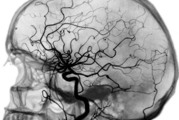
 results Patients often that appeal to the neuropathologist with various complaints, the doctor advised to undergo a diagnostic method as angiography - what is it and how is it conducted? Angiography of cerebral vessels - this is one of the methods of X-ray examination, which allows to determine:
- where and how much there was a blockage of an artery;
- presence and localization of abnormal vasodilation;
- in which part of the brain there was a hemorrhage;
- degree of spread of the tumor.
What is Angiography
vessels Asked what angiography of brain vessels, it is necessary to understand the technique of the procedure. This inspection method involves intravenous radiopaque substance with further visualization of the vascular bed with a special technique( X-ray apparatus, a computer, a magnetic resonance imaging).In this case, angiography allows you to see in what state all the vessels are: not only arteries, but also lymphatic ducts, as well as veins. Typically
cerebrovascular angiography is used to diagnose:
- atherosclerosis( cholesterol plaques appearance);
- stroke( ischemic, hemorrhagic);
- aneurysm;
- cyst formations;
- tumors;
- shunts( non-physiological messages between the vessels).
In addition, angiography of cerebral arteries is prescribed before any surgical interventions on the organs of the nervous system.
 You know how to simultaneously examine the vascular system of the head and neck. Methods of diagnosis.
You can find out all about CT scan here.
How is angiography performed?
Understanding what angiography is, let's find out how this procedure is carried out. Diagnostic manipulation is performed by an angiosurgeon in a hospital setting. To begin with, the doctor punctures( inserts a needle or catheter) the chosen artery, usually a carotid, vertebral column. Then the procedure is performed in several stages:
- Introduction of a radiopaque iodine-containing substance that poorly passes R-rays.
- X-ray head photography in lateral and direct projections( for better visualization, the doctor takes a few shots).
- After passing the contrast on the arteries and getting it into the venous blood, the pictures are repeated again to assess the state of the venous system of the brain.
- Extraction of the puncture needle or catheter.
Please note! During the procedure, the patient can feel a rush of heat, a burning face and a bright blush, a taste of metal in his mouth.
On average, the procedure takes no more than 60 minutes. After it, the patient remains under observation for up to 8 hours, for which possible complications and side effects are excluded. Depending on the device used to visualize blood vessels, release:
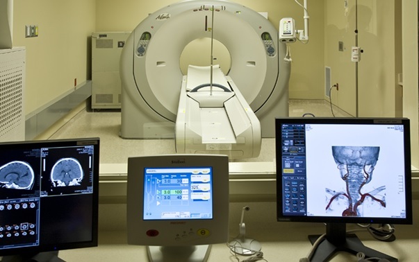
- CT angiography of cerebral vessels - method, linking classical angiography and the opportunity to layered images of good quality. Due to this, a more detailed image appears that allows not only to estimate the size and localization of the vessel, but also its relationship to other arteries and veins. Also with the help of CT diagnostics it is possible to determine the parameters of the blood flow in the vessels under study.
-
 MSCT angiography of cerebral vessels - diagnostic manipulation using a multispiral computer tomograph. Without compromising image quality, the procedure is carried out quickly and with a minimum of radiation exposure.
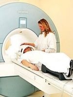
 - MR angiography of cerebral vessels is a study that is performed using the action of a magnetic field. Modern technologies allow obtaining high-precision and bright images, visualized in 3d-projection.
- Selective angiography - in contrast to the general, in selective angiography contrasts only part of the vessels of the brain.
Preparation for examination
Angiography of cerebral vessels, the cost of which varies from 3000 to 10,000 rubles, requires some preparation:
- before the test, a test for susceptibility to radiopaque substance is mandatory( the doctor injects 2 ml of the drug and watches the patient's response within an hour).When unwanted symptoms appear - skin itch, rash, nausea, severe headache - the procedure will have to be abandoned;
- angiography is performed strictly on an empty stomach - the last meal should be 8-10 hours before the study;
- before the procedure, it is necessary to remove all metal ornaments( including hairpins, hairpins) and extractable dentures.
 Find out what is MRI of cerebral vessels. Indications for MRI.
All about MRI of the brain read here.
In which cases it is necessary to find the CT at this address: http: //golmozg.ru/diagnostika/ provedenie-kt-golovnogo-mozga-v-moskve.html. Opportunities for research.
Evaluation of results
When examining images, the doctor evaluates the number and location of blood vessels, both arterial and venous, the presence of pathological constrictions or enlargements, and communication between vessels. In the medical report, the specialist indicates for which disease the characteristic picture is.
write the question in the form below:


