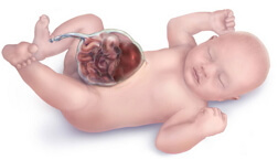
Omphalocele
 Ophalocele is a congenital severe developmental malformation, which manifests itself in the fact that the organs of the abdominal cavity( the liver and intestine) of the fetus come out through the umbilical ring, which is pathologically enlarged. The omphalocele of a newborn is a severe congenital anomaly associated with the highest lethality level of the neonatal period. The problem of the curability of omphalocele lies in the laboriousness of surgical specific correction of the defect, in the high incidence of combined severe congenital anomalies. Despite the severity of the pathology, significant encouraging results of therapy have been achieved, the prognosis of the child's continued viability, a satisfactory future development has improved. The main aspect in nursing such children is the institution where the delivery will take place, ideally, it should be highly specialized centers equipped with high-tech surgical, neonatal resuscitation.
The omphalocele of the fetus - what is it?
Omphalocele occurs in 2-3 children per 10 000 live births. Diagnosis of this defect, at the stage of modern possibilities of prenatal diagnosis, is not difficult. But there are false positive results, often associated with the fact that the ophalocele adopts a physiological "hernial" protrusion of intestinal loops in the fetus. It occurs during the rotation( rotation) of organs, which is a normal process of development of the digestive tract. At the same time, the abdominal cavity is small and the peritoneum is not fully formed, therefore, for the normal development of the intestine and its rotation, part of the loops leave through the "weak" place, and after completion of all processes it returns to its place and is covered by the formed peritoneum. This stage of development occurs at the time from 9 to 14 weeks of gestation. It is during this period and there are flaws in the diagnosis of omphalocele and gastroschisis( the exit of the intestine and stomach through a defective opening in the anterior wall of the abdomen).Therefore, the diagnosis of omphalocele is more reliable during examination, starting from the 15th week of pregnancy.
Ultrasonic picture of omphalocele displays visualization of the intestine in the region of the umbilical ring that extends beyond the abdominal cavity. If there is a suspicion of having an omphalocele when examined at a women's center, the ultrasound specialist, together with the doctor who leads the pregnant woman, decides whether to refer the woman to the prenatal commission. The perinatal consultation includes: obstetrician-gynecologist, geneticist and neonatologist, cardiologist, children's surgeon and resuscitator. Given the high probability of associated vices of vital systems, the possibility of their curability, the viability of the newborn, the surgical options and the volume of resuscitation care, the tactics of conducting the current pregnancy, the nature of the method of labor, the forecast of disability, the adequacy of the development of the child in the future are chosen.
The omphaloceles of the newborn and fetus are rarely found in isolated form. Very often this anomaly is a component of the set of symptoms characteristic of a particular chromosomal disease. More than 20 syndromes are described in the scientific literature, where omphalocele is mandatory. The most common of these are: trisomy 18 pairs( Edwards syndrome) and trisomy 13 pairs( Patau syndrome), Beckwith-Wiedemann syndrome and trisomy 21 pairs( Down syndrome).Congenital anomalies, with a high incidence, that are combined with an omphaloceles are:
- Cloacal exstrophy( abdominal wall defect, with the exit of the bladder divided in two by the large intestine, outwards);
- Pentad of Cantrell( defect of the thoracic wall, pericardial defect( serosa of the heart), heart disease with its exit into the defect of the chest wall, diaphragmatic hernia, omphalocele);
- Syndrome of amniotic cords( filaments formed from the walls of the bladder that entangle body parts and cause various defects);
- Anomalies in the development of the body's stalk( malformation of the wall of the body from the side of the abdomen, accompanied by the exit of the abdominal cavity to the outside, anomaly of the position of the heart, vertebral and limb defects).
In the management plan of pregnancy, burdened by the presence of fetal omphalocele, a mandatory item is the consultation of a geneticist, with the carrying out of karyotyping( quantitative and qualitative chromosome recruitment).Invasive( penetrating into the body) studies can be conducted as:
- Amniocentesis( puncture of the wall of the bladder for sampling of amniotic( amniotic) water);
- Chorion biopsy( sampling for the analysis of the chorion tissues( external envelope of the fetus) through the cervical canal);
- Cordocentesis( through the puncture of the wall of the bladder the fetal umbilical cord is sampled).
On the basis of the data obtained and the decision of the consultation, the specialists decide the feasibility and possibility of maintaining a pregnancy. If, on the conclusion of the prenatal commission, the pregnancy continues, the method of delivery is chosen individually in each case, if the size of the omphalocele is small, then natural births are allowed. But, in most cases, the fetus is removed by caesarean section.
Ophalocele: causes of
The exact cause, as well as the factor causing the omphalocele of the newborn and fetus, is not currently found. The most likely occurrence of a defect at the time of the rotation during the turn of the intestine - the obligatory and physiological stage in the development of the digestive tract. When the torque is disturbed, the "physiological" umbilical hernia is preserved, which, combined with a defect in the development of the peritoneum, forms an omphalocele. There is a theory that this pathology can occur even at the stage of laying and formation of embryonic tissues, from which in the future organs and components of the abdominal wall will form.
Ophalocele belongs to embryo-fetopathies, that is, it develops during the embryonic period( the first three months of pregnancy).Therefore, the first trimester, the so-called "critical" period, is the most important and responsible period for fetal development.
factors that trigger the development of various anomalies, malformations and fetal malformations:
- extragenital( unrelated diseases genital organs) pregnancy pathology: diseases of the respiratory and cardiovascular systems, endocrinopathies, genetic and chromosomal abnormalities, blood disorders, oncopathology, less growth150 cm;
- drug addiction, addiction to alcohol, smoking as a future mother, and father;
- burdened obstetrical history: young age( under 18 years), age primigravida( 35 years), recurrent miscarriage and birth of small for gestational age children, infertility from any cause, death of children during the prenatal, childbirth and early neonatal( first 7 days)Period, cases of the birth of children with malformations;
- reception of teratogenic( fetus-causing) drugs: lithium salts, Warfarin, Colchicine, Aminopterin, Quinine, Thalidomide, etc.;
- current obstetric complications of pregnancy( early severe toxemia and threat of interruption, intrauterine infection, pregnancy-induced and immunological conflict etc.;
- effect on pregnant harmful environmental factors( domestic, industrial chemical toxins, radiation, exposure to pathological temperature, noise and vibration).
Ophalocele: symptoms and signs
The clinical manifestation of omphaloceles in a newborn is the exit of the contents of the abdominal cavity through the pathologically widenedNoe umbilical ring. Size omphalocele determines which bodies eventriruyut( beyond) from the hernial ring. At a small defect( 5 centimeters) and secondary( 10 centimeters) in the umbilical ring located intestinal loops, with a large defect( 10 centimeters anda.) - bowel loops and a portion of the liver if the size of omphalocele large and has the diaphragm defect, the hernia sac can get heart
hernial omphalocele bag presented membranes umbilical cord, the edges are umbilical vessels( two arteries and Vienna) and converge at the top of the hernia. Protrusion, forming an umbilical cord of a normal structure. According to the shape of the hernial protrusion, the omphalocele can be spherical, hemispherical, mushroom-shaped.
If there is a rupture of the hernial sac, then the evolved organs look like in gastroschisis: the intestinal wall is edematous, covered with a layer of fibrin.
omphalocele and gastroschisis are similar in appearance and both these vices are the vices of the anterior abdominal wall, but there are some differences between them, which lead to different tactics when choosing a therapy. With omphaloceles, as with any hernia, there are hernial gates and a sac, and with gastroschisis these components are absent. When gastroschizise never liver event. With omphaloceles, severe defects of other organs and systems are almost always present, and gastroschisis, as a rule, is an isolated defect.
An important point is that with a small defect, a small part of the intestine can enter the ring of the umbilical cord, so it can be taken as a thickened umbilical cord. As a result of this error, the wall of the intestine can be dissected during the treatment of the umbilical cord remnant when the staple or ligature is applied, causing bleeding, necrosis( necrosis of the tissues), and peritonitis( inflammation of the peritoneum).In disputable cases( thick umbilical cord, wide umbilical cord, varicose dilated vessels), the umbilical cord is bandaged 10-15 centimeters away from the base, radiographs are necessarily performed in the projection on the side. If there is an omphalocele on the roentgenogram, the intestinal loops that are outside the abdominal cavity are visible.
Ophalocele is often included in such a severe anomaly as Beckwith-Wiedemann syndrome. For this syndrome, in addition to omphalocele are characteristic: macroglossia( an increase in the tongue that can impede breathing) and an increase in internal organs( pancreas, liver, spleen).Clinically, this syndrome is manifested by hyperinsulinism( increased insulin levels - a hormone that lowers blood sugar levels) and hypoglycemia( low blood sugar).Hypoglycemia is a dangerous condition for a newborn, due to the negative effect of glucose deficiency on brain cells, causing severe encephalopathy. Therefore, in the presence of Beckwith-Wiedemann syndrome, blood glucose monitoring is especially carefully monitored.
Given the combined omphalocele of a newborn with severe defects, it is mandatory to perform additional instrumental techniques. X-rays, neurosonography, echocardiography, ultrasound are carried out.
Ophalocele: operation and treatment of the
The omphalozole of a newborn requires the provision of emergency primary care in the ancestral hall. When a child is born with such a blemish, it is necessary to observe the thermal regime, the child is wiped with a dry diaper, reads the oropharynx according to the indications, decompresses the gastric tube, carries out primary emergency resuscitation if required, and undergoes intubation according to indications with transfer to auxiliary artificial ventilation. The position of the child should be on his side or abdomen. Hernial protrusion is placed in a sterile plastic bag. Transportation to the intensive care unit is carried out in a prepared transport vehicle. In spite of the fact that children with omphalocele, in the majority, donated them put in kuvez, where the most comfortable conditions are created, the optimal parameters of humidity and temperature are selected.
If the size of the omphalocele is very large or if there are multiple combined severe developmental anomalies, then conservative therapy is performed. It consists in the daily processing of the umbilical shells with tanning substances. For this, the omphalocele is fixed vertically in a suspended state and 5% potassium permanganate or Povidone-iodine is applied to the umbilical cord. Over time, as the shells dry up, scar tissue forms and eventually forms a ventral hernia( exit under the skin of the abdominal cavity, through the pathological opening in the anterior wall of the abdomen).
For small and medium-sized omphalocele, a radical one-stage surgical procedure is performed. In this case, an important condition is that the volume of the hernia should correspond to the capacity of the abdominal cavity. The operation consists in carefully adjusting the organs, not creating high intra-abdominal pressure, observing the peculiarities of the correct arrangement of organs, which was broken in the unfinished turn at the embryonic stage of development. After this, all layers of the anterior abdominal wall are layer-by-layer. The operation is terminated by the formation of a "cosmetic" navel. If an omphalocele of small size is combined with a persistent yolk duct, the operation is supplemented by its resection( excision).
For large sizes of ophalocele, stage surgical care is used. The first stage opens the hernial sac and a silicone sterile bag with silastic coating is sutured to the fascial layer of the abdominal wall, and, as far as possible, the organs are fed. Then the bag is tied and fixed in a suspended state vertically. In the course of an average of two weeks, the organs gradually self-correcting and the package is methodically tied up at the bottom. The second stage removes the silicone bag and a small ventral hernia forms from the skin flap of the abdomen. The last stage of surgical treatment will be the correction of the ventral hernia at the age of 6 years, with careful suturing of the abdominal wall layers and the obligatory formation of the "cosmetic" navel.
After surgery, an important moment is a prolonged parenteral nutrition( bypassing the digestive tract), which is carried out through deep venous lines. To do this, use special preparations for intravenous administration, containing the optimal composition of proteins and carbohydrates, fats, various vitamin supplements, microelements, electrolytes. Long-term infusion therapy with the introduction of maintenance drugs, antibacterial therapy, bleeding prophylaxis, etc. is carried out.
Respiratory( respiratory) therapy is performed by assisting artificial ventilation, to alleviate the condition of the child and to prevent overexpiration of the stomach and intestines by air.
Correction of congenital heart defects, blood vessels, brain, urinary and bone systems is performed according to vital indications by specialists of a narrow profile( cardiologists, orthopedists, neurosurgeons, angiologists, urologists, etc.).
Ophalocele: consequences of
Consequences of such a defect as an omphalocele may be as follows:
- Rupture of the umbilical cord. If this occurs in utero, an aseptic( without infectious agent) inflammation of the organs occurs, as a result of which they are covered with a fibrin membrane. If the rupture occurs after birth, the inflammation will be caused by the attachment of a secondary microflora;
- Sepsis. A terrible complication that develops as a result of the entry into the blood of a pathogen from the primary focus, followed by a total spread of the infection;
- Bleeding. Occurs when the umbilical cord is ruptured, traumatized by the intestinal wall, liver, mesentery vessels;
- Necrotizing the intestinal wall. Disturbance of blood supply, drying, inflammation promotes necrosis of intestinal tissues;
- Intestinal adhesive obstruction. The main reason for the formation of intestinal obstruction is the formation of connective tissue adhesions between the intestinal loops, inside the lumen and other organs of the abdominal cavity.
- Invalidation due to the need for superposition of entero-or colostomy, which significantly reduces the quality of life.
Lethal outcome, disability due to isolated omphalocele is practically not found. The cause of the unfavorable outcome are the combined severe malformations and anomalies of the organs.
Omphalocele is a congenital severe defect, the diagnosis of which is possible during the intrauterine stage, which allows you to determine the plan of the present pregnancy and the tactics regarding the future of the newborn. The main problem of operative correction of this anomaly lies in the fact that isolated omphalocele is extremely rare. Damage to the set and quality of chromosomes, severe vices of vital internal organs are the main companions of omphalocele. It is the curability of the combined pathology and is the fundamental prediction of life, disability, further development.
The level of development of perinatal care allows to adequately help such children, and in some cases provides the corresponding functionality of the organism, contributing to the possibility of a full-fledged quality life.


