False joint after fracture: causes and treatment
The process of bone splicing after fracture is characterized by the formation of "bone callus", which is a mass that has no clear forms and structure( high looseness).To make the bones become more precise, doctors use different methods - for example, applying gypsum, applying metal plates or spokes for reliable alignment of fragments / fragments, stretching the bones of the skeleton and so on.But even with such a competent approach to the treatment of fractures, there are cases when the tubular bone simply does not grow together.The result is a smoothing of the contiguous edges of the bone and the formation of a false joint - in medicine this formation is called pseudoarthrosis.
In general, the considered complication of fractures is considered quite common - if a patient is diagnosed with a closed fracture of bone, doctors predict the development of a false joint with a probability of 5-11%, but with open - in 8-35% .Most often, the pathology under consideration takes place with a fracture of the femoral neck, a little less often - with a fracture of the radius, and if this pathology is congenital in nature - on the shin.
Table of contents: Causes of false joints Classification of false joints Symptoms of false joint( pseudoarthrosis) Diagnostic measures Treatment of false jointCauses of false joint
The appearance of an inborn false joint is always associated with any intrauterine fetal pathology.This kind of the considered pathological condition is, in fact, quite rare - for 190,000 newborns, only one case. The reasons for the birth of a child with a false joint can be:
- fibrous dysplasia;
- neurofibromatosis Recklinghausen;
- amniotic constriction;
- embryonic defect of blood vessels due to their underdevelopment.
Acquired false joints - a common complication of fractures and their causes are clearly defined by the physicians:
- consequences of surgical interventions - for example, incorrectly performed fixation of bone fragments when there is no necessary strength of the joint, or resection;
- purulent complications of fractures;
- improper treatment of fractures - for example, a patient too early began to load a limb, or the doctor was forced several times during the treatment to change the cast;
- improperly performed immobilization of the damaged limb with gypsum, violation of the rules of skeletal traction, early removal of the apparatus for fixing fragments;
- some diseases that can lead to disruption of normal regeneration of bones and metabolism - rickets, tumor cachexia, general intoxication of the body, pathology of the endocrine system.
In addition to the foregoing, there are several provoking factors that can also lead to the appearance of the acquired false joint:
- presence of a large number of fragments / bone fragments;
- injuries to the periosteum by medical personnel during various procedures;
-
 long-term use of anticoagulants or steroid medications;
 - ingress of soft tissues or foreign bodies into the gap between bone fragments;
- previously diagnosed with osteoporosis;
- inadequate response of the body to metal devices during the metalloesteosynthesis, when the bone is fixed with plates, bolts and nails;
- obtaining additional tissue damage along with a fracture - for example, irradiation or burns;
- violation of rules for matching the ends of a broken bone;
- no hematoma between the ends of the broken bone;
- clogging and closing by the plate of the bone marrow canal in fragments;
- circulatory disturbance in the area of the location of fragments;
- large distance between the ends of the broken bone.
Classification of false joints
Depending on what was the provoking factor or the true cause of the condition under consideration, distinguish congenital and acquired pseudoarthrosis.If we consider this pathology from the side of the nature of the lesion, then only fire and non-fire-resistant pseudoarthroses will be isolated. But the classification of false joints according to their clinical manifestations is more detailed:
- The forming false joint.Occurs at the end of the period, which is necessary for normal bone growth.X-rays help to identify clear boundaries of the "gap" of the fracture and the bone callus.The patient complains of constant drawing pain in the area of formation of a false joint, and when trying to touch it indicates an increase in the intensity of pain.
- Fibrous pseudarthritis.The doctor clearly diagnoses the presence of fibrous tissue located between the bone fragments, and the result of the X-ray will be a clearly defined gap between them.With such a false joint, if it is formed in the region of the joints, the mobility of the latter becomes sharply limited.Necrotic false joint.It occurs often after wounds of a gunshot nature, but it can also be in fractures if there is a high probability of bone necrosis.Similar purulent pseudoarthrosis doctors are often diagnosed with injuries of the neck of the talant and femoral or mid-part of the scaphoid bone.
- Pseudoarthritis of bone regenerate.He appears with an incorrect osteotomy of the tibia, if the doctor has violated the rules of stretching or poorly performed fixation with the use of special equipment for lengthening the segments.
- True false joint( neoarthrosis).Most often it develops on single-bone segments with excessive mobility.Similar pseudoarthrosis is characterized by the formation on the edges of bone fragments of fibrous cartilaginous tissue with areas of hyaline cartilage.Around the wreckage appears an education, which in its composition and appearance resembles a periarticular bag.
By the method of formation and intensity of bone formation the considered pathological condition is classified as follows:
- hypertrophic false joint - the bone tissue begins to grow particularly at the ends of the damaged bone;
- normotrophic false joint - no breaks are found on bone fragments;
- atrophic false joint - blood flow is inadequately determined, insufficient bone formation, osteoporosis can be diagnosed.
In addition, a false joint can be uncomplicated - a condition in which there is no infection or the appearance of pus at the site of pseudoarthrosis. But in some cases, doctors diagnose "infected pseudoarthrosis", which means that a purulent infection has been connected.In this case, the patient will have a fistula and a cavity of different sizes at the site of injury, from which the purulent contents of the are periodically released.Most often in such false statutes there are fragments of shells or metal locks.
Symptoms of false joint( pseudoarthrosis)
The signs of the pathological condition under consideration are quite specific, therefore the diagnosis is not difficult.The most pronounced symptoms of a false joint are:
- increasing the amplitude of movements, changing their orientation, which can not be called characteristic for the limb;
- slightly below the fracture site, a large edema with clear boundaries is formed;
- atypical mobility of those parts of the body in which there should be no normal movements;
- changes in the functions of joints located close to the site of the fracture;
- muscles of the limb lose their characteristic strength - with a false joint the patient can not squeeze fingers, raise an easy object;
- violation of the functions of the fractured limb.
Diagnostic measures
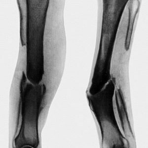
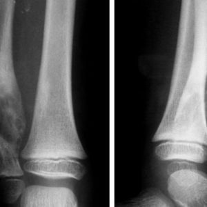
A completely informative diagnostic method for suspected false joint formation is conventional radiography.Computer tomography is extremely rare, only in the case of a severe course of fracture and an unclarified false joint.
The study of X-ray images with pseudoarthrosis helps the physician identify:
-
 unconnected fragments, that is, a bone callus characteristic for fractures is absent;
 - fragments of broken bone become rounded and smooth;
- infection and the appearance of end plates at the ends of fragments of the bone cavity, which can stop regeneration in the tissues of the bone marrow;
- gap located between the "articular surfaces".
X-ray images allow only to reveal and confirm the fact of the presence of a false joint, but to determine the degree of bone formation and diagnose the specific form of the pathology in question, the patient will be assigned a radioisotope study.
Treatment of a false joint
The main method of treatment of a considered pathological condition is surgical operation .The goal of such treatment is to restore the continuity of the broken bone and then the doctors take measures to eliminate deformities.The tactics of treatment are selected individually, because it all depends on the specific clinical case and the characteristics of the patient's body.
False joint is eliminated by general and local treatment.
General medical measures
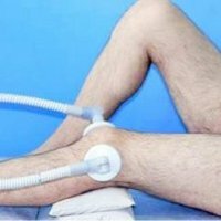
By this term I mean activities that are aimed at increasing muscle tone, normalizing blood circulation directly at the site of formation of a false joint, doctors try to keep the functionality of the damaged lower or upper limb as much as possible.To achieve these goals, the patient is assigned various physiotherapy procedures, massage and a set of exercises for the treated gymnastics.
Local treatment
Implies the operation, the purpose of which is to create favorable conditions for the fusion of bone fragments.During the work with the patient, the surgeon not only restores the normal shape of the bone by bringing the fragments together and connecting them, but also ensures normal blood circulation in this place .Preventive measures that are aimed at preventing infection and the development of purulent inflammation are considered to be mandatory in this case.
Local treatment is carried out by different methods:
- bone plastic;
- compression-distraction osteosynthesis;
- is a stable osteosynthesis.
The specific tactics of local treatment are chosen depending on the type of false joint.For example, if it has a hypertrophic form, then a compression-distraction apparatus will simply be applied to the limb.But with atrophic pseudoarthrosis will have to carry out bone plastic surgery.
Compression-distraction osteosynthesis3
This method of treatment involves the use of special apparatus that will provide a comparison of bone fragments.The physician must ensure complete immobility of the injured limb, and already in this state, the use of the apparatus begins, which will approximate and combine the fragments of the bone.It is compression-distraction osteosynthesis that helps specialists to eliminate shortening and / or deformation of the extremities.
Stable osteosynthesis
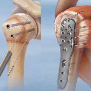 This method of treating a false joint involves the use of metal parts( plates or rods) that will ensure the fusion of damaged bone.To impose them, the surgeon will have to completely expose the bone at the site of the fracture - this operation is performed under general anesthesia.
If the patient is diagnosed with hypertrophic pseudoarthrosis, then surgery on bone plastic surgery is not required, but in case of treatment of atrophic pseudoarthrosis it is necessary.
Bone plastic
Seldom, before the operation, it is necessary to eliminate any inflammation and make sure there are no cicatrical changes.If any, first purulent inflammation is cured and excision of cicatricial changes is performed. Bone plastic surgery can be performed only 8 months after the indicated treatment, but doctors usually survive 12 months.
If a false joint is treated, the affected limb should be immobilized( immobilized) for a fairly long time.Once physicians are allowed to move, the patient must undergo a course of restorative therapy.In the framework of this rehabilitation period, massage and physical therapy courses are provided, physiotherapy procedures, spa treatment can be prescribed.
In general, the result of such a complex treatment is usually excellent - in 72% of cases, patients were discharged home with completely restored functions of the injured limb .
False joint is a pathology that is very easy to diagnose, therefore doctors recommend simply to undergo a full course of treatment, which will be appointed by the attending physician - treatment in any case will be timely.
Tsygankova Yana Aleksandrovna, medical reviewer, therapist of the highest qualification category