Iridocyclitis: causes, symptoms, treatment, complications, prognosis
Content
- general information
- ICD code
- Anatomy of the eye
- Causes and classification
- Iridocyclitis symptoms
- Complications
- Diagnostics
- Treatment of iridocyclitis
- Forecast
general information
Iridocyclitis (anterior uveitis) is an inflammatory disorder that affects certain structures in the eye, in particular the iris and ciliary body. In most cases, the cause is unknown and most often affects young people.
Iridocyclitis is the most common among uveitis, and its causes are recognized:
- eye injury;
- viral and bacterial infections;
- systemic diseases, especially autoimmune diseases;
- allergic reactions;
- diabetes.
Clinically, iridocyclitis manifests itself with symptoms such as:
- eye pain and profuse lacrimation;
- redness and burning of the eyes;
- photophobia, i.e. hypersensitivity to light;
- blurred vision.
For diagnosis, history and physical examination are used with blood tests and instrumental tests. Recognizing and treating iridocyclitis requires the experience of an ophthalmologist (ophthalmologist).
Iridocyclitis is an important pathology that requires immediate treatment in order to avoid the development of complications, which, if very severe, can seriously damage the structures of the eye to the point of causing blindness.
Treatment is etiological, i.e. aims to eliminate the root cause. Typically, corticosteroid and mydriatic drugs (which dilate the pupil) are used to reduce signs of inflammation and minimize the risk of serious complications.
ICD code
ICD 10 - Iridocyclitis (H20)
Anatomy of the eye
Speaking about the anatomy of the eye, we can say that it essentially consists of three layers:
- the sclera and cornea, which represent the outer layer;
- uvea, which represents the middle choroid, that is, the one that contains the blood vessels necessary to support all ocular structures;
- retina - the inner lining of the eye, which is the peripheral part of the visual analyzer.
Uvea is divided into 3 parts:
- iris (iris);
- ciliary (ciliary) body;
- choroid (choroid).

Based on the anatomy of the eye, iridocyclitis therefore corresponds to anterior uveitis or inflammation of the so-called anterior chamber of the eye (which includes the iris and ciliary body).
The iris forms the anterior part of the choroid and separates the anterior cornea from the lens from behind. The iris is the structure responsible for eye color due to the presence of various types of pigments in its deepest layer.
Read also:Allergic conjunctivitis
Inside the iris is the pupil, a veritable "hole" that allows light to enter the eye and thus can reach the retina from behind.
The function of the iris is to regulate the amount of light that must reach the retina, depending on the ambient lighting conditions.
- In bright light, the pupil constricts (miosis) due to the contraction of the constricting muscle of the pupil.
- Conversely, in conditions of low luminosity, the pupil dilates (mydriasis) due to the dilating muscle of the pupil, allowing more light to reach the retina.
The ciliary body is probably a circular structure that compresses with the lens and performs the function of accommodation. This term refers to the ability of the human eye to focus on objects at different distances: ciliary the body, by contracting or unclenching, changes the curvature of the lens, allowing the object to be focused for several minutes.
Causes and classification
Inflammation of the iris and ciliary body can have a clinical course:
- acute when it comes on suddenly and has a limited duration;
- chronic, when iridocyclitis persists and recurs for at least 3 months.
In addition, it can be:
- unilateral if it affects only one eye;
- bilateral, less often.
The main causes of iridocyclitis are:
- idiopathic, the most common cause (not associated with any recognizable systemic disease);
- postoperative uveitis;
- eye injury;
- diabetes;
- allergic reactions;
- adverse drug reactions (very rare);
- as a result of an autoimmune disease, a large group of diseases that include:
- arthritis:
- rheumatoid arthritis;
- psoriatic arthritis;
- juvenile idiopathic arthritis;
- lupus;
- ankylosing spondylitis;
- sarcoidosis;
- brucellosis;
- Behcet's disease;
- inflammatory bowel disease (Crohn's disease and nonspecific ulcerative colitis);
- Whipple's disease;
- arthritis:
- infections:
- Virus:
- herpes simplex;
- shingles;
- cytomegalovirus;
- HIV;
- Bacteria:
- staphylococci or streptococci;
- tuberculosis;
- syphilis;
- leprosy;
- Parasites:
- toxoplasmosis;
- toxocariasis;
- Mushrooms:
- candidiasis;
- histoplasmosis;
- Virus:
- primary ocular syndromes:
- Fuchs syndrome;
- sympathetic ophthalmia;
- glaucoma;
- primary or secondary cancer of the eye.
Iridocyclitis symptoms

Symptoms that characterize iridocyclitis:
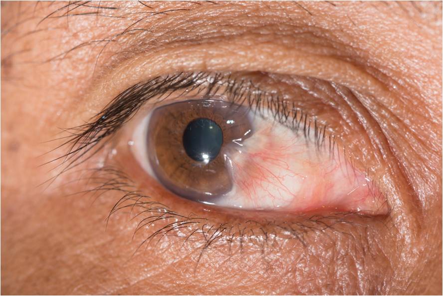
- red eyes, with inflammatory conjunctival hyperemia;
- pain in the eyes, mono or bilateral, depending on the degree of the process;
- photophobia, i.e. hypersensitivity to light, which becomes very annoying, sometimes painful;
- profuse lacrimation;
- burning and irritation;
- visual disturbances such as decreased visual acuity or blurred vision;
- permanent miosis, i.e. constriction of the pupil;
- opacity of the cornea;
- blepharospasm, tonic and spastic contraction of the eyelids.
Read also:Abrasions and scratches on the cornea of the eye
Complications
Iridocyclitis can lead to rare but serious complications, especially when there is an evolution towards the panuveitis form:
- glaucoma (increased intraocular pressure, which over time damages the retina and can lead to blindness);
- retinal detachment;
- inflammation of the cornea, sclera and other ocular structures by expanding the inflammatory process;
- secondary cataract (lens opacity).
Diagnostics
The diagnosis of iridocyclitis begins with a history and physical examination:
-
Anamnesis (medical history) consists of the formulation of a number of questions by the doctor, which are necessary to investigate and restore the entire medical history of the patient. In this case, it is important to obtain information about the possible presence of:
- autoimmune disease;
- allergies;
- diabetes;
- previous eye injuries.
-
Physical examination allows you to recognize all the symptoms reported by the patient, and objective clinical signs. At this stage, it is important to make a differential diagnosis with other pathologies similar to iridocyclitis, such as:
- conjunctivitis;
- retinitis pigmentosa;
- heavy keratitis.
To this end, the doctor may perform laboratory tests with blood tests or some instrumental eye examinations, such as:
- slit lamp inspection;
- analysis with a tonometer to determine intraocular pressure;
- examination with an ophthalmoscope, after pupil dilation.
These procedures often allow the doctor to detect some of the clinical signs specific to iridocyclitis, such as:
- blepharospasm;
- Tyndall's phenomenon (the presence of floating cells in aqueous humor);
- nodules at the level of the iris or pupil;
- ocular hypotension, decreased intraocular pressure;
- keratic sediments;
- hypopyon (accumulation of pus in the lower part of the anterior chamber of the eye);
- anterior or posterior synechia between the iris and the cornea, as well as between the iris and the lens (situations with a risk of cataract or glaucoma formation),
An eye biopsy is rarely useful when a small sample of cells is taken at the level and subsequently analyzed by a pathologist under a microscope.
It is important to suspect iridocyclitis in every patient experiencing:
- Pain in the eyes;
- redness;
- photophobia;
- initial decrease in vision.
Read also:Redness of the eyes: causes and treatment
Treatment of iridocyclitis

Treatment for iridocyclitis has three main goals:
- Alleviate pain and discomfort that significantly reduce quality of life.
- Heal the root cause to achieve final healing.
- Avoid and treat any complications before they become serious and irreversible.
From a general point of view, drug treatment involves the use of the following drugs:
- corticosteroids (topical or systemic in severe cases);
- mydriatics (which dilate the pupil) to relieve pain and other inflammatory signs.
Treatment of iridocyclitis must be started early to prevent irreversible damage. For treatment, corticosteroids are almost always prescribed, usually in the form of eye drops. Corticosteroids can also be taken by mouth or injected into the eye or into the tissues around the eye. Also used are drugs that dilate the pupil, such as homatropine or cyclopentolate in droplets.
Drugs may be prescribed to treat specific causes of uveitis. For example, if the cause is an infection, drugs may be prescribed to kill the infectious organisms (antibiotics, antivirals).
In some cases, other treatments are necessary, such as surgery, laser therapy, oral administration, or vein injection (intravenous) of drugs that suppress the immune system (immunosuppressants) if autoimmune is the cause disease.
A blindfold can help in the most acute phase of the disease.
Forecast
In general, the prognosis is good for uncomplicated iridocyclitis, especially when it has a resolvable infectious basis. More problems arise with complex iridocyclitis, such as those associated with autoimmune diseases, which tend to become chronic and therefore require long-term treatment.
If left untreated, anterior uveitis can quickly destroy the eye. The disease can cause long-term, vision-threatening symptoms such as corpus luteum edema, retinal damage and cataracts.