Autoimmune myositis: symptoms, treatment, diagnosis, prognosis
Content
- What is autoimmune myositis?
- Causes of autoimmune myositis
- Symptoms and Signs
- Diagnostics
- Autoimmune myositis treatment
- Forecast
What is autoimmune myositis?
Autoimmune myositis Is a group of autoimmune rheumatic diseases that cause inflammation and muscle weakness (polymyositis) or skin and muscle (dermatomyositis).
These diseases lead to muscle inflammation (myositis), muscle weakness causing disability, and sometimes soreness. Weakness is usually seen in the shoulders and hips, but it can also affect the symmetrical muscles of the body.
Causes of autoimmune myositis
Autoimmune myositis usually develops in adults between the ages of 40 and 60, or in children between the ages of 5 and 15.
Women are twice as likely to develop it as men. In adults, the disease can occur independently or in a complex of connective tissue diseases, for example, with a mixed connective tissue disease, systemic lupus erythematosus or systemic sclerosis.
The cause of autoimmune myositis has not been established. Viruses or autoimmune reactions may be involved in the development of the disease. Cancer can also cause this disorder. It is possible that an immune response directed against a malignant tumor may also be directed to components of muscle tissue. This disease can be hereditary.
There are four types of autoimmune myositis:
- Polymyositis.
- Dermatomyositis.
- Necrotizing immune-mediated myopathies.
- Myositis with inclusions.
Dermatomyositis usually causes skin changes that do not occur with polymyositis, which helps in the differential diagnosis of both diseases. Muscle tissue biopsies also look different under a microscope.
Necrotizing immune-mediated myopathies are diseases in which muscle cells (myocytes) die, but tissues other than muscle are not affected.
Inclusion myositis is a separate disease with symptoms similar to those of chronic polymyositis of unknown etiology. However, the disease develops in the elderly and often affects other muscles (for example, muscles of the hands and stop), has a longer course, responds poorly to treatment, and the muscles have a different appearance under microscope.
Symptoms and Signs
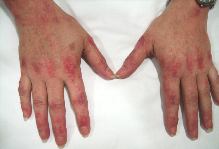
The symptoms of autoimmune myositis are similar in people of all ages, but it turns out that muscle inflammation often develops more suddenly in children than in adults. Symptoms that may start during or immediately after an infection include:
- Symmetrical muscle weakness (especially in the upper arms, thighs and lower legs);
- joint pain (although mild muscle pain is more common);
- difficulty swallowing;
- heat;
- fatigue and weight loss.
It can also occur Raynaud's phenomenon, which is characterized by sudden blanching and tingling sensations in the fingers, or numbness in response to exposure to cold or emotional distress.
Read also:Periarteritis nodosa
Muscle weakness may develop slowly or suddenly, with a gradual worsening over weeks or months. Due to the fact that the lesion primarily affects the muscles near the center of the body, serious difficulties with movements such as raising an arm above shoulder level, walking up stairs, and lifting from a chair or seat toilet bowl. If the muscles in the neck are affected, even lifting the head off the pillow may become impossible. Individuals with shoulder or hip muscle weakness may require the use of a wheelchair or constant bed rest. Injury to the muscles in the upper esophagus can lead to difficulty swallowing and regurgitation of food. In this case, muscle damage to the hands, feet and face usually does not occur.
Joint pain and inflammation present in about 30% of patients. Pain and swelling are mostly minor.
Internal organs usually not affected, except for the throat and esophagus. However, damage to the lungs and heart is not excluded, leading to disturbances in the heart rhythm (arrhythmias), shortness of breath and cough. Symptoms of damage to the gastrointestinal tract, which may occur in children but are usually absent in adults, are caused by inflammation of the blood vessels (vasculitis). Such symptoms can include bloody vomiting, black, tarry stools, and severe abdominal pain, sometimes with a hole (perforation) in the lining of the intestine.
People with dermatomyositis have skin changes. A rash usually occurs along with muscle weakness and other symptoms. A dark or purple rash appears on the face, with reddish purple puffiness around the eyes (a symptom of "purple glasses"). The rash can be bulging and flaky and occur on almost any part of the skin, but is most common on the knuckles, elbows, knees, on the outer side of the upper thighs and partially on the hands and stop. Redness and hardening of the skin around the nails is possible.
When the rash disappears, brownish discoloration, scarring, wrinkling, or depigmented pale patches may appear on the skin. A rash on the scalp can resemble psoriasis and itch a lot. Sensitivity to sunlight and skin ulcers are also observed. Bumps may develop under the skin or in muscle tissue, especially in children, due to calcium deposits. Raised reddish bumps may appear in the metacarpophalangeal joints (called Gottron papules) and sometimes in the interphalangeal joints.
Sometimes these characteristic skin changes are observed in the absence of muscle weakness and inflammation. In this case, the disease is called amyopathic dermatomyositis.
Diagnostics
The following criteria are used to diagnose autoimmune myositis:
- muscle weakness in the shoulders or hips and legs;
- sometimes a characteristic rash;
- Elevated blood levels of certain muscle enzymes (especially creatine kinase), indicating muscle damage
- pathological changes in the electrical activity of muscles based on the results of electromyography or changes in the appearance of muscles in images obtained using magnetic resonance imaging (MRI);
- characteristic changes in muscle tissue found on biopsy and observed under a microscope (the most reliable confirmation).
Read also:Symptoms and treatment of Reiter's syndrome (disease)
Muscle biopsy is often prescribed and is the most reliable way to diagnose autoimmune myositis, especially in doubtful cases. Other laboratory tests cannot unequivocally identify autoimmune myositis, but may help rule out other disorders, detect the risk of complications and determine the severity diseases.
Blood tests are used to measure levels of antinuclear antibodies (ANA) and other antibodies that are present in most people with autoimmune myositis. Although blood test results can help diagnose autoimmune myositis, they alone cannot confirm the diagnosis. autoimmune myositis, since the abnormalities detected with their help are sometimes present in healthy people or in people with other diseases. The diagnosis of autoimmune myositis is made based on all the information the doctor has gathered, including symptoms, results of a physical examination, and results of all examinations.
MRI can also help you select a biopsy site. To rule out other muscle disorders, samples of muscle tissue are taken for specific examinations.
Doctors often order cancer screening for people age 40 and older who have dermatomyositis, or among people aged 60 and older suffering from polymyositis, since such patients may have hidden malignant tumors.
Autoimmune myositis treatment

Often, the condition of patients is favorably affected by a moderate restriction of physical activity during periods of the most severe manifestations of muscle inflammation.
Typically assigned prednisone (a corticosteroid) for oral administration in high doses. This drug slowly increases muscle strength, relieves pain and swelling, while maintaining disease control. For people with severe illness with difficulty swallowing or weakness of the respiratory muscles, intravenous corticosteroidssuch as methylprednisolone. Many adult patients are forced to continue taking low-dose prednisone or an alternative a drug for many years or even an indefinite period, for prophylaxis relapse.
To monitor the disease's response to corticosteroid treatment, doctors periodically do blood tests to measure muscle enzyme levels. Levels usually decline to normal or near-normal, and muscle strength returns after 6-12 weeks. An MRI can also detect areas of inflammation, which allows the doctor to determine the response of the disease to the prescribed treatment. Once enzyme levels return to normal, the dose of prednisone can be gradually reduced. As the levels of muscle enzymes increase, the dose is increased.
Read also:Behcet's disease
Although corticosteroids are usually given first when treating people with autoimmune myositis, these drugs cause side effects (for example, high blood sugar, mood swings, cataracts, risk of fractures and glaucoma), especially when given in high doses and for a long time. Therefore, prednisone may be supplemented with prednisone to shorten the duration of corticosteroid use and minimize side effects. immunosuppressants (such as methotrexate, tacrolimus, azathioprine, mycophenolate mofetil, rituximab, or cyclosporine). As another method of treatment, immunoglobulin (a substance containing various antibodies in large quantities) may be given to be injected into a vein (intravenously). Some patients receive a combination of corticosteroids, immunosuppressants, and immunoglobulin.
If muscle weakness is due to cancer, the response to prednisone treatment is usually low. However, the severity of this condition is usually alleviated if it is possible to effectively treat the malignant tumor.
Patients receiving corticosteroids are at increased risk of fractures due to osteoporosis. To prevent osteoporosis, these patients are prescribed drugs used to treat osteoporosis, such as bisphosphonates, vitamin D and calcium supplements. People receiving immunosuppressants are also prescribed drugs to prevent infections caused by fungi such as Pneumocystis jirovecii.
Patients with autoimmune myositis are at increased risk of developing atherosclerosis and are under close medical supervision.
Forecast
Up to 50% of patients (especially children) who received treatment within 5 years after diagnosis, often reach a long period of asymptomatic course (remission), and some may even completely get well. However, a return (relapse) of the disease is possible at any time. Once diagnosed, approximately 75% of patients have a minimum life expectancy of 5 years. This indicator is even higher among patients of childhood.
In adults, the risk of death is increased due to severe and progressive muscle weakness, difficulty swallowing, malnutrition, inhalation of food and subsequent development pneumonia (aspiration pneumonia) or respiratory failure, which often develops simultaneously with pneumonia.
Children with juvenile dermatomyositis may develop severe inflammation of the blood vessels (vasculitis), the blood supply to the intestine, which, if untreated, can ultimately lead to intestinal perforation.
The course of polymyositis is characterized by greater severity and resistance to treatment among patients with lesions of the heart and lungs. Polymyositis and especially dermatomyositisare associated with an increased risk of developing cancer. In patients with malignant diseases, the main cause of death is the tumor, not autoimmune myositis.