Radiation diagnosis of the bone system
The human body is a mystery and we are striving to solve it. Modern medicine has stepped far ahead. Now research and the identification of a disease is not difficult. You can explore everything, starting with the surface of the skin, the internal organs and ending with bones. Today we will talk about the study of the human skeleton.
Methods of radiation examination of the bone system
Radiation diagnosis is a group of methods that allow to see the structure, diseases and pathologies of internal organs, but it acts non-invasively, i.e. Without affecting the skin of needles or any surgical instruments.
The following diagnostic methods are used to study the bone system.
Ultrasound diagnosis
This is the safest method of investigation. It gives the most complete information about the state of the body, does not harm with frequent use. The method does not require special preparation of the body for research and does not take much time. Used to study the state of the musculoskeletal system to detect damage to tendons, nerve endings, blood vessels, muscles, and the detection of old and acute joint injuries.
X-ray densitometry
The method that allows using X-ray radiation to determine the mineral density of the bone tissue of the spine, the necks of the hip bones. This method is unique in the radiodiagnosis of osteoporosis at an early stage for the detection of fractures. Indications for the use of this method are broken bones and spine, pain in the joints, spine and bones, endocrine system diseases, menopause and the period after breast-feeding. The method can not be used for critical days and pregnancy.
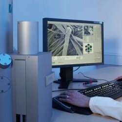
Computer tomography
This is a method of diagnosing the bone system, in which X-ray images of the desired parts of the skeleton are obtained in a layered form. At the same time, deviations from norms are detected. This method is used in the study of the head, spine, joint-bone system, as well as organs of the abdominal cavity and chest to detect injuries and tumors. The method is contraindicated in menstruation and pregnancy.
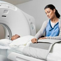
Magnetic resonance imaging( MRI)
The method allows to perform bone system examination without using a radial load. This is a non-invasive method, safe and reusable without any consequences. In MRI, contrast agents are used to more fully perform the diagnosis of the area of interest. The method is used for diseases of the spine, bones, joints, as well as organs of the chest and peritoneum for the detection of tumors, traumatic injuries and tumor-like diseases. Contraindicated in claustrophobia, menstruation, the presence of pacemakers and implants with metal parts, in the early stages of pregnancy.
Classical X-ray diagnostics
This is the leading method of studying the bone system, and is used to detect skeletal injuries, a variety of bone pathologies and diseases of the musculoskeletal system. This method is contraindicated in pregnancy.
The tasks of this method are:
- Determining the type and nature of the fracture.
- Detection of a dislocation.
- Disclaimer or confirmation of a previous diagnosis.
- Detection of foreign bodies.
- Controlling the healing of fractures.
- Finding the degree of displacement of debris and their number.
- Confirmation of correctness of treatment.
Methods of radiation diagnostics help:
- To study the occurrence of physiological processes in the joints and bones.
- Identify injuries and various diseases.
- Assess the body's relation to various factors of mechanical, toxic effects, etc.
- Watching the construction of the skeleton.
- To talk about the restructuring of an organism of a pathological and functional nature.
- To assess the morphological state of the bone system( ie outlines of bones, their shape, density, structure, etc.).
Radiation diagnosis is very useful for medicine due to its accuracy, speed, painlessness and the ability to more fully examine the state of the bone system of the body.