US of the testicles: when is it conducted and what allows to reveal?
The testicular ultrasound is a noninvasive examination of the scrotum organs. This safe and informative examination makes it possible to evaluate the structure, the size of the testicles, and to reveal many pathologies.
Contents of
- 1 What diseases can detect ultrasound testicular?
- 2 Indications for
- 3 Contraindications to
- 4 procedure Preparation of
- 5 Procedure for
- 6 Decoding of results: testicular dimensions
What diseases can detect ultrasound of the testicular?
Ultrasound examination of the scrotum organs( in particular, the testicles) makes it possible to determine the presence of the following pathologies:
- Hydrosele. is a collection of fluid in the area of one or two testicles. Otherwise, called dropsy testicles. A hydrocele can be congenital, and can be acquired as a result of an inflammatory process.
- Varicocele. Varicocele is a varicose veins of the testicles. Usually bilateral. In addition to diaphanoscopy, mandatory ultrasound is indicated.
- Infertility. In itself, infertility can only be determined by laboratory methods, but ultrasound can show abnormalities in the structures of testicles, which can be the reason for the inability to conceive a child.
- Tumors of testicles. Both benign and malignant. The disadvantage of the method is the inability to determine the nature of the neoplastic process. To verify the diagnosis, a diagnostic puncture( biopsy) is shown, followed by a histological and morphological study.
- Cysts of testicles and appendages. Cysts are hollow sacs filled with exudate: intercellular fluid, blood, pus, etc.
- Injuries. First of all, doctors are interested in the consequences of the injured testicles.
- Abscesses. Simply put "abscesses".In their echogenic structure they differ from both tumors and cysts, so ultrasound is practically a non-alternative diagnostic method.
- Epididymitis. Otherwise - inflammation of the epididymis.
These diseases are most common in the practice of urologists and urologists andrologists.
Indications for
There are clear indications for ultrasound testing of the testicles:
-
testicular ultrasound reveals common urological pathologies.
If there is an established and verified spermogram diagnosis of "infertility."Or if the diagnosis is not yet worth, but a married couple can not conceive a child for a year or longer.
- If urological operation is to be performed. Especially on the testicles.
- With an increase in the testes, appendages, which may indicate inflammation.
- If there is a suspicion of inguinal hernia. Moreover, when there is reason to suspect a partial loss of abdominal organs in the scrotum.
- When there are persistent problems with normal erection, while the indicators of laboratory tests( including hormones) are within normal limits.
- In the absence of testicles or both testicles.
- When there is swelling of the scrotum for no reason.
- A similar study is assigned to monitor the dynamics of the development of diseases, including tumors, chronic pathologies.
- With suspicions of orchitis and other inflammatory lesions of the testicles and appendages.
- There are suspicions of varicose veins testicles.
- Urgently conducted in case of injury, development of hematoma.
- To assess the condition of the testicles in inflammation of the regional lymph nodes. This may indicate the onset of the neoplastic process.
- When suspected of a tumor process.
- If the adolescent is experiencing too early or, alternatively, later puberty.
This is not an exhaustive list of indications. In any doubtful cases, the doctor has every reason to assign an ultrasound testicular. There are also additional indications, if we are talking about boys under the age of majority:
- for suspected hypogonadism( a lack of sex hormone deficiency when working glands).
- If there is reason to suspect congenital diseases of the testicles.
- Often in newborns and small children, the testicle does not fall independently. In this case, ultrasound is designed to identify the root cause of the problem.
Contraindications to procedure
Why there are no contraindications:
- This method of research is non-invasive, that is, it does not interfere with the work of organs and body systems.
- Ultrasonic waves do not have any negative impact on the body. There is not one proven fact of causing harm.
- Such a study provides an opportunity to assess not only the structure of tissues, but also the speed, as well as the nature of the blood flow.
Accordingly, this is an informative, but safe method of diagnosis. In addition, painless, because you can recommend it even to children.
Preparation of
No specialized training is required. The only exception is children. The child needs to talk about the procedure, explain the absence of painful sensations, i.e. Prepare mentally. With a need to take a diaper or a towel to wipe the gel after carrying out ultrasound.
Procedure of
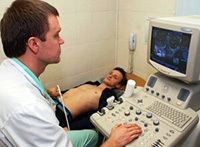
The method is based on the effect of ultrasound on the tissue. Ultrasound is reflected from dense bodies, including body tissues, giving a clear picture. This is called the principle of echolocation.
The ultrasound procedure is quite typical for any localization. The doctor puts the patient on his back or side. The scrotum is lubricated with a special gel, after which the procedure begins. The doctor drives the sensor and records the results. It is absolutely painless and does not require any effort from the patient. The whole examination lasts about 10-20 minutes, in exceptional cases, the duration of the study can be extended to half an hour for a more detailed study. After that, the examinee can return to their daily business
Decoding results: indicators of testicular size
decipher the results of the research is impossible without specialized medical education. This should deal with an andrologist or urologist. However, it is simply necessary to know some of the indicators for the patient himself. The main indicator is the size of the testicles. In adult men, the size of the testicles varies between 40-60 mm in length( 4-6 cm) and 25-30 mm in width( 2.5-3 cm).In newborns, the size of the testicles is within 15 mm in length and 10 mm in width. The expanded data are presented in the table. Age
| left testicle size( width / length in cm) | right testicle size( width / length in cm) | |
|---|---|---|
| 2-8 years | 0.76 / 1.53 | 0.78 / 1.48 |
| 8-10 years | 0.88 / 1.82 | 0.86 / 1.82 |
| 11-13 years | 1.62 / 3.12 | 1.62 / 3.09 |
| 14-17 years | 2.05 / 3.96 | 2.12 / 3.84 |
Ultrasound has a lot of advantages, and in some cases there are no alternatives available. It allows you to identify a variety of diseases. Therefore, one should not be afraid of such a study.
See also: Why and how to perform a testicle massage?
Recommended for viewing: